

Effects of valsartan on ventricular arrhythmia induced by programmed electrical stimulation in rats with myocardial infarction
- PMID: 22128836
- PMCID: PMC3823086
- DOI: 10.1111/j.1582-4934.2011.01502.x
Effects of valsartan on ventricular arrhythmia induced by programmed electrical stimulation in rats with myocardial infarction
Abstract
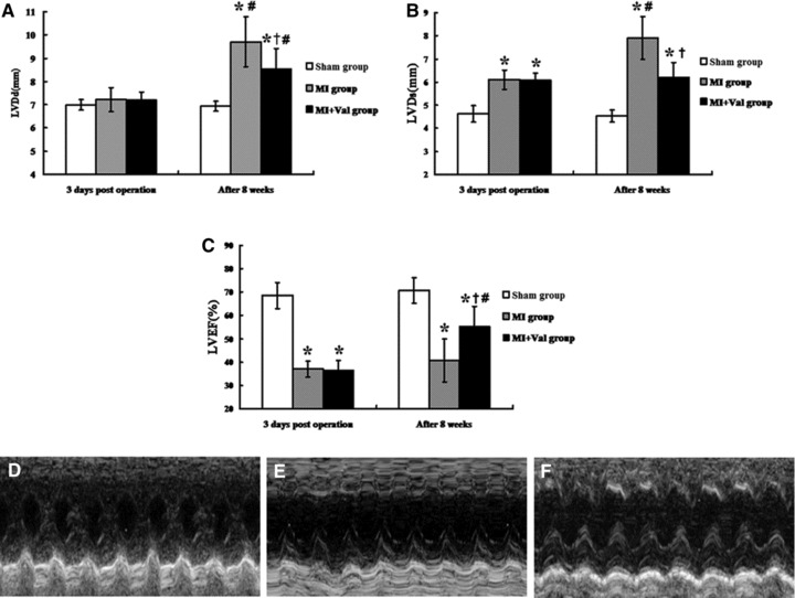
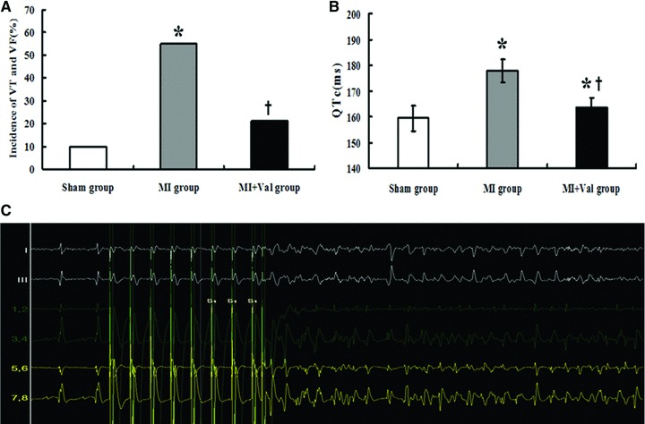
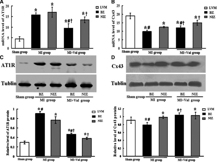
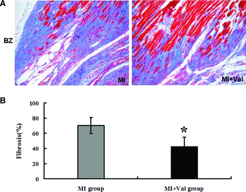
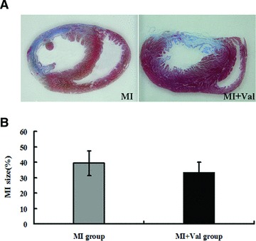
The impact of angiotensin II receptor blockers (ARBs) on electrical remodelling after myocardial infarction (MI) remains unclear. The purpose of the present study was to evaluate the effect of valsartan on incidence of ventricular arrhythmia induced by programmed electrical stimulation (PES) and potential link to changes of myocardial connexins (Cx) 43 expression and distribution in MI rats. Fifty-nine rats were randomly divided into three groups: Sham (n = 20), MI (n = 20) and MI + Val (20 mg/kg/day per gavage, n = 19). After eight weeks, the incidence of PES-induced ventricular tachycardia (VT) and fibrillation (VF) was compared among groups. mRNA and protein expressions of Cx43, angiotensin II type 1 receptor (AT1R) in the LV border zone (BZ) and non-infarct zone (NIZ) were determined by real-time PCR and Western blot, respectively. Connexins 43 protein and collagen distribution were examined by immunohistochemistry in BZ and NIZ sections from MI hearts. Valsartan effectively improved the cardiac function, reduced the prolonged QTc (163.7 ± 3.7 msec. versus 177.8 ± 4.5 msec., P < 0.05) after MI and the incidence of VT or VF evoked by PES (21.1% versus 55%, P < 0.05). Angiotensin II type 1 receptor expression was significantly increased in BZ and NIZ sections after MI, which was down-regulated by valsartan. The mRNA and protein expressions of Cx43 in BZ were significantly reduced after MI and up-regulated by valsartan. Increased collagen deposition and reduced Cx43 expression in BZ after MI could be partly attenuated by Valsartan. Valsartan reduced the incidence of PES-induced ventricular arrhythmia, this effect was possibly through modulating the myocardial AT1R and Cx43 expression.
© 2012 The Authors Journal compilation © 2012 Foundation for Cellular and Molecular Medicine/Blackwell Publishing Ltd.
Figures
Similar articles
-
[On angiotensin II receptor distribution after myocardial infarction in dogs].Zhonghua Xin Xue Guan Bing Za Zhi. 2009 Apr;37(4):358-62. Zhonghua Xin Xue Guan Bing Za Zhi. 2009. PMID: 19791474 Chinese.
-
Combination renin-angiotensin system blockade and angiotensin-converting enzyme 2 in experimental myocardial infarction: implications for future therapeutic directions.Clin Sci (Lond). 2012 Dec;123(11):649-58. doi: 10.1042/CS20120162. Clin Sci (Lond). 2012. PMID: 22715807
-
Upregulation of angiotensin II type 2 receptor and limitation of myocardial stunning by angiotensin II type 1 receptor blockers during reperfused myocardial infarction in the rat.J Cardiovasc Pharmacol Ther. 2003 Sep;8(3):217-26. doi: 10.1177/107424840300800307. J Cardiovasc Pharmacol Ther. 2003. PMID: 14506547
-
Valsartan in the treatment of heart attack survivors.Vasc Health Risk Manag. 2006;2(2):125-38. doi: 10.2147/vhrm.2006.2.2.125. Vasc Health Risk Manag. 2006. PMID: 17319456 Free PMC article. Review.
-
Enhancing cardiac protection after myocardial infarction: rationale for newer clinical trials of angiotensin receptor blockers.Am Heart J. 2000 Jan;139(1 Pt 2):S23-8. doi: 10.1067/mhj.2000.102904. Am Heart J. 2000. PMID: 10618584 Review.
Cited by
-
Progression of infarct-mediated arrhythmogenesis in a rodent model of heart failure.Am J Physiol Heart Circ Physiol. 2021 Jan 1;320(1):H108-H116. doi: 10.1152/ajpheart.00639.2020. Epub 2020 Nov 8. Am J Physiol Heart Circ Physiol. 2021. PMID: 33164577 Free PMC article.
-
Valsartan prevents gefitinib-induced lung inflammation, oxidative stress, and alteration of plasma metabolites in rats.Saudi J Biol Sci. 2023 Feb;30(2):103522. doi: 10.1016/j.sjbs.2022.103522. Epub 2022 Dec 1. Saudi J Biol Sci. 2023. PMID: 36561332 Free PMC article.
-
Cardiac ablation of Rheb1 reduces sodium currents in infant mice.Int J Clin Exp Med. 2014 Apr 15;7(4):947-54. eCollection 2014. Int J Clin Exp Med. 2014. PMID: 24955166 Free PMC article.
-
Angiotensin II type 1 receptor blockade attenuates gefitinib-induced cardiac hypertrophy via adjusting angiotensin II-mediated oxidative stress and JNK/P38 MAPK pathway in a rat model.Saudi Pharm J. 2022 Aug;30(8):1159-1169. doi: 10.1016/j.jsps.2022.06.020. Epub 2022 Jun 22. Saudi Pharm J. 2022. PMID: 36164571 Free PMC article.
-
Angiotensin II Receptor Blockers and Arrhythmias in Ventricular Hypertrophy.J Am Heart Assoc. 2022 Aug 2;11(15):e026634. doi: 10.1161/JAHA.122.026634. Epub 2022 Jul 19. J Am Heart Assoc. 2022. PMID: 35862170 Free PMC article. No abstract available.
References
-
- Brezins M, Elyassov S, Elimelech I, et al. Comparison of patients with acute myocardial infarction with and without ventricular fibrillation. Am J Cardiol. 1996;78:948–50. - PubMed
-
- Pascale P, Schlaepfer J, Oddo M, et al. Ventricular arrhythmia in coronary artery disease: limits of a risk stratification strategy based on the ejection fraction alone and impact of infarct localization. Europace. 2009;11:1639–46. - PubMed
-
- Nakamura Y, Yoshiyama M, Omura T, et al. Beneficial effects of combination of ACE inhibitor and angiotensin II type 1 receptor blocker on cardiac remodeling in rat myocardial infarction. Cardiovasc Res. 2003;57:48–54. - PubMed
-
- Pfeffer MA, McMurray JJ, Velazquez EJ, et al. Valsartan, captopril, or both in myocardial infarction complicated by heart failure, left ventricular dysfunction, or both. N Engl J Med. 2003;349:1893–906. - PubMed
Publication types
MeSH terms
Substances
LinkOut - more resources
Full Text Sources
Medical
