. 2011 Nov 30:8:167.
doi: 10.1186/1742-2094-8-167.

Lipopolysaccharide-enhanced transcellular transport of HIV-1 across the blood-brain barrier is mediated by luminal microvessel IL-6 and GM-CSF

Affiliations

Lipopolysaccharide-enhanced transcellular transport of HIV-1 across the blood-brain barrier is mediated by luminal microvessel IL-6 and GM-CSF

Shinya Dohgu et al. J Neuroinflammation. .

Abstract

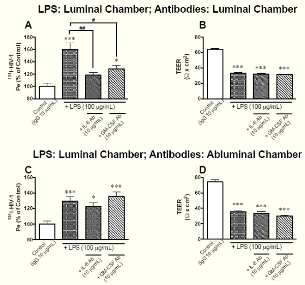
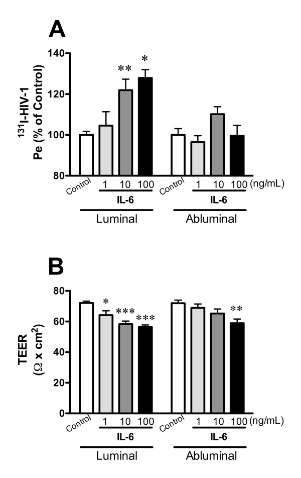
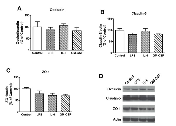
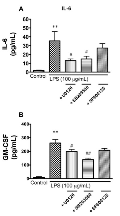
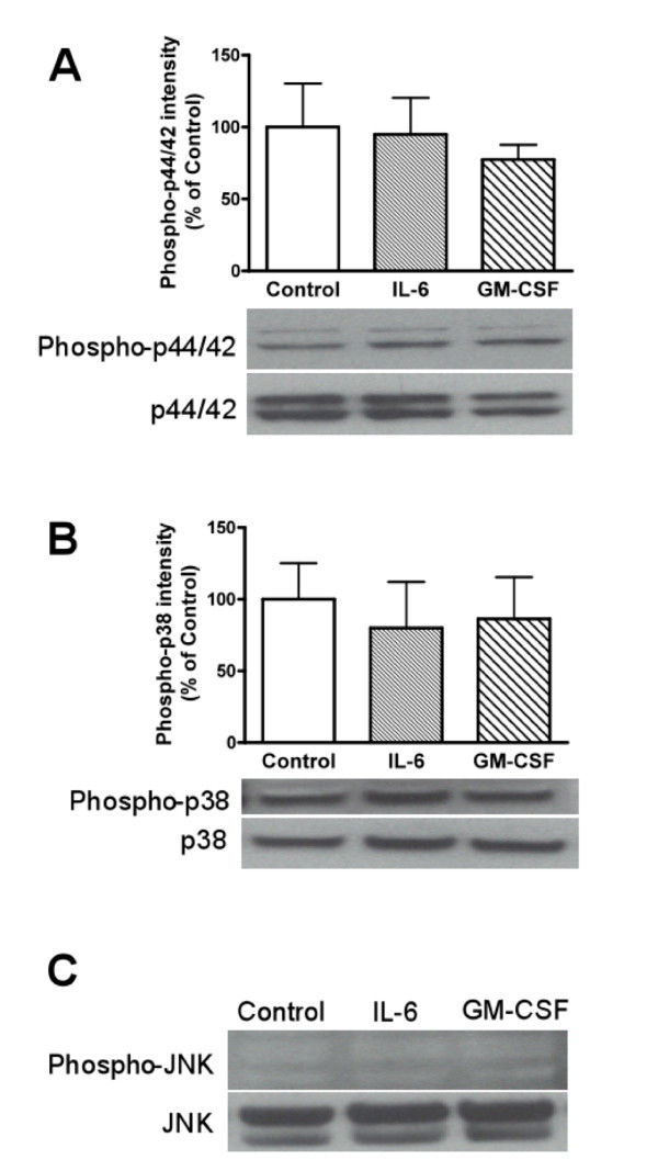
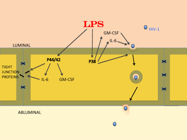
Elevated levels of cytokines/chemokines contribute to increased neuroinvasion of human immunodeficiency virus type 1 (HIV-1). Previous work showed that lipopolysaccharide (LPS), which is present in the plasma of patients with HIV-1, enhanced transcellular transport of HIV-1 across the blood-brain barrier (BBB) through the activation of p38 mitogen-activated protein kinase (MAPK) signaling in brain microvascular endothelial cells (BMECs). Here, we found that LPS (100 μg/mL, 4 hr) selectively increased interleukin (IL)-6 and granulocyte-macrophage colony-stimulating factor (GM-CSF) release from BMECs. The enhancement of HIV-1 transport induced by luminal LPS was neutralized by treatment with luminal, but not with abluminal, antibodies to IL-6 and GM-CSF without affecting paracellular permeability as measured by transendothelial electrical resistance (TEER). Luminal, but not abluminal, IL-6 or GM-CSF also increased HIV-1 transport. U0126 (MAPK kinase (MEK)1/2 inhibitor) and SB203580 (p38 MAPK inhibitor) decreased the LPS-enhanced release of IL-6 and GM-CSF. These results show that p44/42 and p38 MAPK signaling pathways mediate the LPS-enhanced release of IL-6 and GM-CSF. These cytokines, in turn, act at the luminal surface of the BMEC to enhance the transcellular transport of HIV-1 independently of actions on paracellular permeability.


Figures

Figure 1
Figure 1
Effects of antibodies to IL-6 and GM-CSF on LPS-induced changes in the permeability of BMECs to 131I-HIV-1 (A and C) and TEER (B and D). LPS (100 μg/mL) was added to the luminal chamber. In panels A and B, antibodies to IL-6 or GM-CSF were added to the luminal chamber. In panels C and D, antibodies to IL-6 or GM-CSF were added to the abluminal chamber. After 4 hr of incubation, transport study was performed. In panels A and C, results are expressed as % of control. Control values were 1.52 ± 0.16 × 10⁻⁵ and 1.52 ± 0.05 × 10⁻⁵ cm/min (A and C, respectively). Values are means ± SEM (n = 9-15). *P < 0.05, ***P < 0.001, significant differences from control. #P < 0.05, ##P < 0.01, significant differences from LPS (100 μg/mL).
Figure 2
Figure 2
Functional polarity to IL-6 in BMEC permeability of HIV-1 (A) and TEER (B). BMECs were exposed to IL-6 (1, 10, and 100 ng/mL) in the luminal or abluminal chamber for 4 hr. In panel A, results are expressed as % of control. The control values of permeability coefficient for 131I-HIV-1 in panel A was 1.03 ± 0.11 × 10⁻⁵ and 1.07 ± 0.08 × 10⁻⁵ cm/min for the luminal and abluminal control, respectively. Values are means ± SEM (n = 3-12). *P < 0.05, **P < 0.01, ***P < 0.001, significant difference from each corresponding control.
Figure 3
Figure 3
Functional polarity to GM-CSF in BMEC permeability of HIV-1 (A) and TEER (B). BMECs were exposed to GM-CSF (1, 10, and 100 ng/mL) in the luminal or abluminal chamber for 4 hr. In panel A, results are expressed as % of control. The control values of permeability coefficient for 131I-HIV-1 in panel A was 137 ± 0.13 × 10⁻⁵ and 1.32 ± 0.13 × 10⁻⁵ cm/min for the luminal and abluminal control, respectively. Values are means ± SEM (n = 3-12). **P < 0.01, significant difference from control.
Figure 4
Figure 4
Effects of LPS, IL-6, and GM-CSF on the expression of tight junction proteins in BMECs. BMECs were exposed to LPS (100 μg/mL), IL-6 (100 ng/mL), or GM-CSF (100 ng/mL) for 4 hr. Expression levels of occludin, claudin-5, ZO-1, and actin were detected by western blot. Relative intensity of occludin (A), claudin-5 (B), and ZO-1 (C) is calculated as ratio of arbitrary densitometric units of target protein to that of actin. Results are expressed % of control. Values are means ± SEM (n = 3). (D) Photographs are representative in three independent experiments.
Figure 5
Figure 5
Effects of various MAPK inhibitors on LPS-enhanced release of IL-6 (A) and GM-CSF (B) by BMECs. BMECs were treated with LPS (100 μg/mL) for 4 hr in the presence or absence of U0126 (10 μM), SB203580 (10 μM), or SP600125 (10 μM). Values are means ± SEM (n = 5-8). **P < 0.01, significant differences from control. #P < 0.05, ##P < 0.01, significant differences from LPS (100 μg/mL).
Figure 6
Figure 6
Effects of IL-6 and GM-CSF on phosphorylation of MAPKs in BMECs. BMECs were exposed to IL-6 (100 ng/mL) or GM-CSF (100 ng/mL) for 4 hr. Western blot analyses were performed to detect phosphorylated p44/42 MAPK (A), p38 MAPK (B), and JNK (C) as well as total p44/42 MAPK, p38 MAPK, and JNK. Relative intensity is calculated as ratio of arbitrary densitometric units of phoshorylated protein to that of total protein. (C) For phospho-JNK, sorbitol-treated PC12 cells were used as positive control (data not shown). Results are expressed % of control. Values are means ± SEM (n = 3-4). Photographs are representative in three to four independent experiments.
Figure 7
Figure 7
Schematic of findings. LPS released both GM-CSF and IL-6 through MAPKs p44/42 and p38 pathways. Previous work has shown that LPS-induced tight junction disruption (paracellular permeability) is mediated through the p44/42 MAPK pathway whereas HIV-1 transcytosis is mediated through the p38 MAPK pathway. Tight junction function as measured by TEER but not tight junction protein expression as measured by western blots was influenced by IL-6 (but not GM-CSF) through a site not blocked by antibodies and so assumed to be intracellular. GM-CSF and IL-6 both promoted HIV-1 transcytosis. The transcytotic effects of GM-CSF and IL-6 were mediated through luminal but not abluminal sites that were blocked by antibodies and therefore assumed to be extracellular.

Similar articles

Cited by

References

    1. Gonzalez-Scarano F, Martin-Garcia J. The neuropathogenesis of AIDS. Nature Rev Immunol. 2005;5:69–81. doi: 10.1038/nri1527. - DOI - PubMed
    1. McArthur JC, Haughey N, Gartner S, Conant K, Pardo C, Nath A, Sacktor N. Human immunodeficiency virus-associated dementia: An evolving disease. J Neurovirol. 2003;9:205–221. - PubMed
    1. Eden A, Fuchs D, Hagberg L, Nilsson S, Spudich S, Svennerholm B, Price RW, Geisslen M. HIV-1 viral escape in cerebrospinal fluid of subjects on suppressive antiretroviral treatment. Journal of Infectious Diseases. 2010;202:1768–1769. doi: 10.1086/657343. - DOI - PMC - PubMed
    1. Banks WA, Akerstrom V, Kastin AJ. Adsorptive endocytosis mediates the passage of HIV-1 across the blood-brain barrier: evidence for a post-internalization coreceptor. Journal of Cell Science. 1998;111:533–540. - PubMed
    1. Banks WA, Freed EO, Wolf KM, Robinson SM, Franko M, Kumar VB. Transport of human immunodeficiency virus type 1 pseudoviruses across the blood-brain barrier: role of envelope proteins and adsorptive endocytosis. Journal of Virology. 2001;75:4681–4691. doi: 10.1128/JVI.75.10.4681-4691.2001. - DOI - PMC - PubMed

Publication types

MeSH terms

LinkOut - more resources