

MC3T3-E1 osteoprogenitor cells systemically migrate to a bone defect and enhance bone healing
- PMID: 22129134
- PMCID: PMC3338109
- DOI: 10.1089/ten.TEA.2011.0545
MC3T3-E1 osteoprogenitor cells systemically migrate to a bone defect and enhance bone healing
Abstract
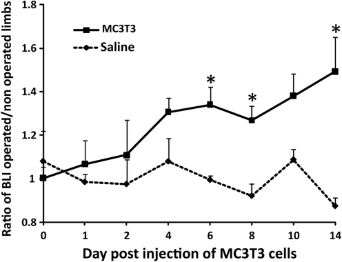
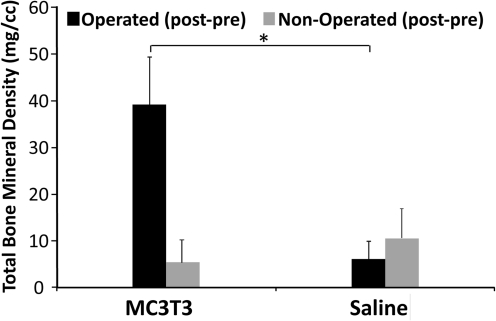
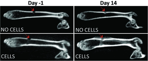
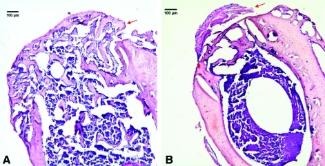
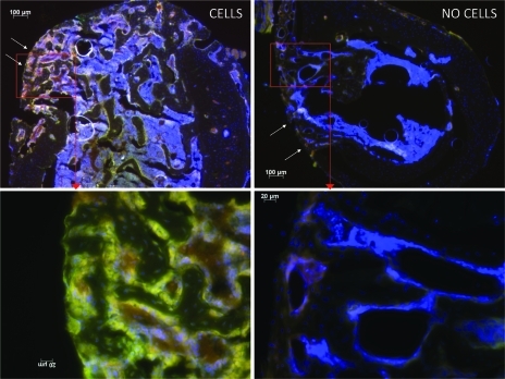
Although iliac crest autologous bone graft remains the gold standard for treatment of bone defects, delayed- and nonunions, and arthrodeses, several alternative strategies have been attempted, including the use of mesenchymal stem cells. Whether cells from the osteoblast lineage demonstrate systemic recruitment to an acute bone defect or fracture, and whether these cells directly participate in bone healing is controversial. This study tests two hypotheses: (1) that exogenous murine MC3T3-E1 osteoprogenitor cells with a high propensity for osteoblast differentiation are able to systemically migrate to a bone defect and (2) that the migrated MC3T3-E1 cells enhance bone healing. Two groups of nude mice were used; a bone defect was drilled in the left femoral shaft in both groups. MC3T3-E1 were used as reporter cells and injected in the left ventricle of the heart, to avoid sequestration in the lungs. Injection of saline served as a control. We used bioluminescence and microCT to assay cell recruitment and bone mineral density (BMD). Immunohistochemical staining was used to confirm the migration of reporter cells. MC3T3-E1 cells were found to systemically migrate to the bone defect. Further, BMD at the defect was significantly increased when cells were injected. Systemic cell therapy using osteoprogenitor cells may be a potential strategy to enhance bone healing.
Figures
References
-
- Mahendra A. Maclean A.D. Available biological treatments for complex non-unions. Injury. 2007;38(Suppl 4):S7. - PubMed
-
- Goulet J.A. Senunas L.E. DeSilva G.L. Greenfield M.L. Autogenous iliac crest bone graft. Complications and functional assessment. Clin Orthop Relat Res. 1997;339:76. - PubMed
-
- Weiss L. Web watch. Tissue Eng. 2002;8:167.
-
- Younger E.M. Chapman M.W. Morbidity at bone graft donor sites. J Orthop Trauma. 1989;3:192. - PubMed
-
- Gerstenfeld L.C. Alkhiary Y.M. Krall E.A, et al. Three-dimensional reconstruction of fracture callus morphogenesis. J Histochem Cytochem. 2006;54:1215. - PubMed
Publication types
MeSH terms
Grants and funding
LinkOut - more resources
Full Text Sources
Other Literature Sources
Medical
