. 2011 Nov 30:11:117.
doi: 10.1186/1472-6750-11-117.

Expression of recombinant multi-coloured fluorescent antibodies in gor -/trxB- E. coli cytoplasm

Affiliations

Expression of recombinant multi-coloured fluorescent antibodies in gor -/trxB- E. coli cytoplasm

Anatoliy Markiv et al. BMC Biotechnol. .

Abstract

Background: Antibody-fluorophore conjugates are invaluable reagents used in contemporary molecular cell biology for imaging, cell sorting and tracking intracellular events. However they suffer in some cases from batch to batch variation, partial loss of binding and susceptibility to photo-bleaching. In theory, these issues can all be addressed by using recombinant antibody fused directly to genetically encoded fluorescent reporters. However, single-chain fragment variable domains linked by long flexible linkers are themselves prone to disassociation and aggregation, and in some cases with isoelectric points incompatible with use in physiologically relevant milieu. Here we describe a general approach that permits fully functional intracellular production of a range of coloured fluorescent recombinant antibodies with optimally orientated VH/VL interfaces and isoelectric points compatible for use in physiological solutions at pH 7.4 with a binding site to fluorophore stoichiometry of 1:1.

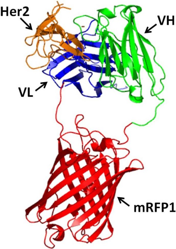
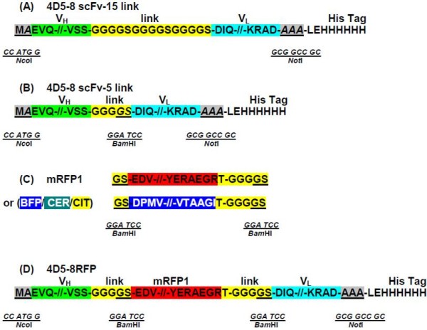
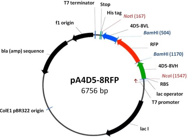
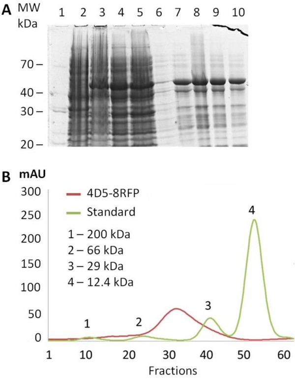
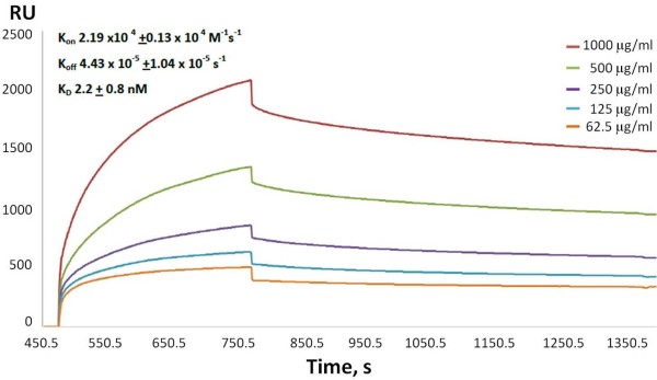
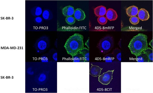
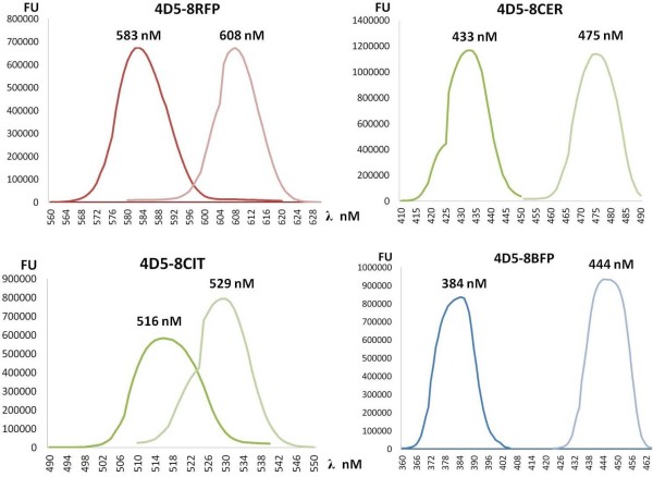
Results: Here we report the design, assembly, intracellular bacterial production and purification of a panel of novel antibody fluorescent protein fusion constructs. The insertion of monomeric fluorescent protein derived from either Discosoma or Aequorea in-between the variable regions of anti-p185HER2-ECD antibody 4D5-8 resulted in optimal VH/VL interface interactions to create soluble coloured antibodies each with a single binding site, with isoelectric points of 6.5- 6. The fluorescent antibodies used in cell staining studies with SK-BR-3 cells retained the fluorophore properties and antibody specificity functions, whereas the conventional 4D5-8 single chain antibody with a (Gly4Ser)3 linker precipitated at physiological pH 7.4.

Conclusions: This modular monomeric recombinant fluorescent antibody platform may be used to create a range of recombinant coloured antibody molecules for quantitative in situ, in vivo and ex vivo imaging, cell sorting and cell trafficking studies. Assembling the single chain antibody with monomeric fluorescent protein linker facilitates optimal variable domain pairing and alters the isoelectric point of the recombinant 4D5-8 protein conferring solubility at physiological pH 7.4. The efficient intracellular expression of these functional molecules opens up the possibility of developing an alternative approach for tagging intracellular targets with fluorescent proteins for a range of molecular cell biology imaging studies.


Figures

Figure 1
Figure 1
A 3D model of the genetically encoded fluorescent antibody. Molecular model (ribbon representation) of the Herceptin™ antigen-binding fragment (Fv) and human Her2 (PDB 1N8Z) complexed with of mRFP1 based on DsRed (PDB 1G7K). A portion of Her2 is shown in orange, VH chain of 4D5-8 in green, mRFP1 in red and VL chain of 4D5-8 antibody in navy blue.
Figure 2
Figure 2
The assembly of the 4D5 recombinant antibody constructs. (A) 4D5-8 scFv 15 amino acid linker. The mature protein begins with VH chain, followed by a (Gly4Ser)3 linker, VLchain. The sites used to directionally insert the in-frame scFv encoding sequence are underlined (NcoI-NotI). (B) 4D5-8 scFv 5 amino acid linker. The mature protein begins with VH chain, followed by a (Gly4Ser), VL chain. The sites used to directionally insert the in-frame scFv encoding sequence are underlined (NcoI-NotI). The linker terminal Gly Ser underlined is encoded by an in frame BamHI site. (C) Modified mRFP1. The mRFP1 sequence is flanked by two in-frame BamHI sites underlined. The alternative BFP, CER and CIT encoding sequences were modified in a similar manner. (D) 4D5-8RFP. The modified mRFP1 (C) was inserted into 4D5-8 scFv 5 amino acid linker construct (B) at the BamHI site.
Figure 3
Figure 3
Plasmid maps of p4D5-8RFP used to clone and express red fluorescent antibody. The expression cassette has a T7 promoter region, a lac operator, a ribosome-binding site followed by a cloning region with NcoI and NotI restriction sites for the insertion of the DNA sequences. At the end, there is a T7 termination sequence to restrict translation to the expression of RNA for the recombinant protein. The vector also has the ampicillin resistance gene, ColE1 pBR322 origin of replication and lacI repressor gene.
Figure 4
Figure 4
Purification and characterization of the 4D5-8RFP recombinant protein. (A) SDS-PAGE and Coomassie stained gel of 4D5-8RFP: molecular markers (Lane 1), uninduced sample (Lane 2), IPTG induced sample (Lane 3), soluble fraction (Lane 4), flow-through Ni2+-nitrilotriacetic acid column (Lane 5), wash fraction (Lane 6), elution fractions using 500 mM imidazole (Lanes 7-10). (B) Gel filtration chromatography of standards proteins (peaks 1-4) and Ni2+-nitrilotriacetic acid enriched 4D5-8RFP protein using Sephadex G 200 gel bead column calibrated with sweet potato b-amylase 200 kDa, bovine serum albumin 66 kDa, bovine erythrocyte carbonic anhydrase 29 kDa and horse heart cytochrome c 12.4 kDa protein standards (Sigma-Aldrich).
Figure 5
Figure 5
Surface plasmon resonance analysis of purified 4D5-8RFP binding to recombinant p185HER2-ECD antigen (SinoBiological Inc, Beijing, China).
Figure 6
Figure 6
Detection of 4D5-8RFP binding to p185HER-2 -overexpressing SK-BR-3 and non-expressing MDA-MD-231 cells by confocal microscopy. Cells incubated with nuclear stain TO-PRO3, actin label Phalloidin-FITC conjugate and 4D5-mRFP. SK-BR-3 cells were also incubated with TO-PRO3 and 4D5-mCIT.
Figure 7
Figure 7
Spectrophotometric determination of 1 mg/mL of 4D5-8 fluorescent proteins, RFP, CER, CIT and BFP.

Similar articles

Cited by

References

    1. Ledbetter JA, Rouse RV, Micklem HS, Herzenberg LA. T cell subsets defined by expression of Lyt-1,2,3 and Thy-1 antigens. Two-parameter immunofluorescence and cytotoxicity analysis with monoclonal antibodies modifies current views. J Exp Med. 1980;152(2):280–295. doi: 10.1084/jem.152.2.280. - DOI - PMC - PubMed
    1. Urano Y, Asanuma D, Hama Y, Koyama Y, Barrett T, Kamiya M, Nagano T, Watanabe T, Hasegawa A, Choyke PL. et al.Selective molecular imaging of viable cancer cells with pH-activatable fluorescence probes. Nat Med. 2009;15(1):104–109. doi: 10.1038/nm.1854. - DOI - PMC - PubMed
    1. Coons AH, Creech HJ, Jones RN, Berliner E. The Demonstration of Pneumococcal Antigen in Tissues by the Use of Fluorescent Antibody. J Immunol. 1942;45(3):159–170.
    1. Griep RA, van Twisk C, van der Wolf JM, Schots A. Fluobodies: green fluorescent single-chain Fv fusion proteins. J Immunol Methods. 1999;230(1-2):121–130. doi: 10.1016/S0022-1759(99)00131-3. - DOI - PubMed
    1. Lu M, Gong XG, Yu H, Li JY. Cloning, expression, purification, and characterization of LC-1 ScFv with GFP tag. J Zhejiang Univ Sci B. 2005;6(8):832–837. - PMC - PubMed

Publication types

LinkOut - more resources