

Review
doi: 10.1146/annurev.psych.121208.131631.
Fear extinction as a model for translational neuroscience: ten years of progress
Affiliations
- PMID: 22129456
- PMCID: PMC4942586
- DOI: 10.1146/annurev.psych.121208.131631
Item in Clipboard
Review
Fear extinction as a model for translational neuroscience: ten years of progress
Annu Rev Psychol.
2012.
Abstract
The psychology of extinction has been studied for decades. Approximately 10 years ago, however, there began a concerted effort to understand the neural circuits of extinction of fear conditioning, in both animals and humans. Progress during this period has been facilitated by a high degree of coordination between rodent and human researchers examining fear extinction. Here we review the major advances and highlight new approaches to understanding and exploiting fear extinction. Research in fear extinction could serve as a model for translational research in other areas of behavioral neuroscience.
Figures
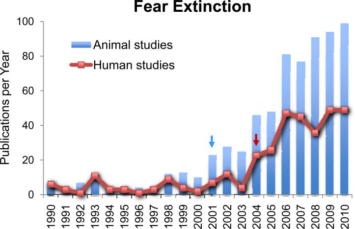
Exponential increase in fear extinction studies within the last decade. Number of peer-reviewed studies identified with key terms “fear” and “extinction” in PubMed, in the past 20 years. Note that the increase in animal studies (blue bars, blue arrow) began several years prior to the increase in human studies (orange line, orange arrow).
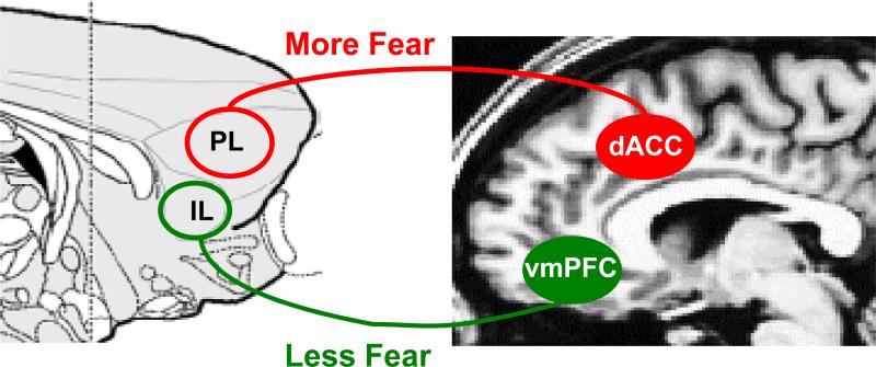
Homologous prefrontal structures in rodent and human brain that modulate fear expression. The rodent prelimbic cortex (PL) and human dorsal anterior cingulate cortex (dACC) increase fear expression and oppose extinction, while the rodent infralimbic cortex (IL) and human ventromedial prefrontal cortex inhibit fear expression and promote extinction.
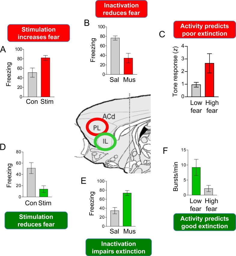
Summary of rodent findings for PL and IL prefrontal cortex. A-C: Divergent findings suggest that PL activity increases fear expression. D-F: Parallel data suggest that IL inhibits fear expression and strengthens extinction recall (D,E,F). Data adapted from previously published studies: A, D: Vidal-Gonzalez et al., 2006: see also (Kim et al 2010); B, E: Sierra-Mercado et al., 2011, see also (Laurent & Westbrook 2009); C: Burgos-Robles et al.,2009; F: Burgos-Robles et al., 2007, see also (Chang et al 2010).
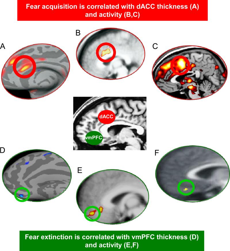
Summary of neuroimaging research demonstrating that the dACC (A-C) regulates fear acquisition, and vmPFC (D-F) regulates fear extinction, in healthy humans. A,B: Milad et al. (2007a), C: Linnman et al. (2011b); D: Milad et al. (2005b), E: Milad et al. (Milad et al 2007b), F: Kalisch et al., 2006.

Prefrontal-amygdala interaction in rodents and humans. A. The tone response of a single PL neuron (blue line, z-score) superimposed upon the rat's freezing to the tone (gray bars). Adapted from Burgos-Robles et al., 2009. Note the high correlation between sustained PL activity and freezing (bin: 3 sec). B. Left: Schematic illustrating reciprocal connections between PL and the BLA. Transient tone response emanating from BLA trigger sustained tone responses in PL that feedback to BLA and drive fear. Right: perievent time histogram showing a conditioned tone response of a PL neuron under control conditions (black bars), and after inactivation of BLA with infusion of muscimol (red bars). Fear signals in BLA drive PL. C. Psychophysiological interaction (PPI) analysis during extinction recall in healthy humans. During recall of a non-extinguished stimulus, increased coupling of the neural activity of the amgydala (seed) with the dACC (red circle) was observed, and reduced functional coupling between the amgydala (seed) and vmPFC (green circle) was observed (adapted from Linnman et al. (2011c).
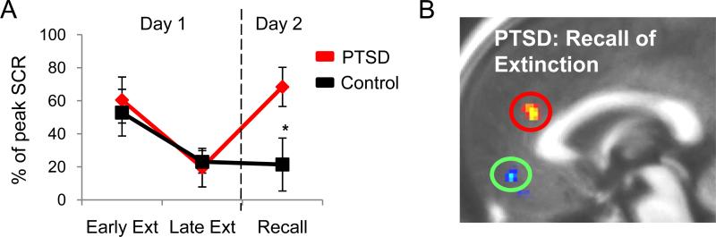
In PTSD, reduced extinction recall is associated with failure to activate vmPFC and increased activation of dACC. A. Skin conductance responses normalized to peak acquisition levels show intact fear learning (indexed in early extinction [early ext]), intact extinction learning (late ext), but impaired extinction recall (day 2) relative to controls. B. fMRI data using a contrast of extinguished vs. unextinguished conditioned stimuli in PTSD vs. Controls during extinction recall, showinge hypoactivation of the vmPFC (green circle) and a trend toward hyperactivation of dACC (red circle) in PTSD relative to controls. Adapted from Milad et al. (2009b)
References
-
- Andreano JM, Cahill L. Sex influences on the neurobiology of learning and memory. Learn. Mem. 2009;16:248–66. - PubMed
Publication types
MeSH terms
Grants and funding
LinkOut - more resources
Full Text Sources
Other Literature Sources
Medical
