Review
. 2012 Feb;24(1):107-15.
doi: 10.1016/j.ceb.2011.10.004. Epub 2011 Nov 28.

Integrin inside-out signaling and the immunological synapse

Affiliations
Review

Integrin inside-out signaling and the immunological synapse

Timothy A Springer et al. Curr Opin Cell Biol. 2012 Feb.

Abstract

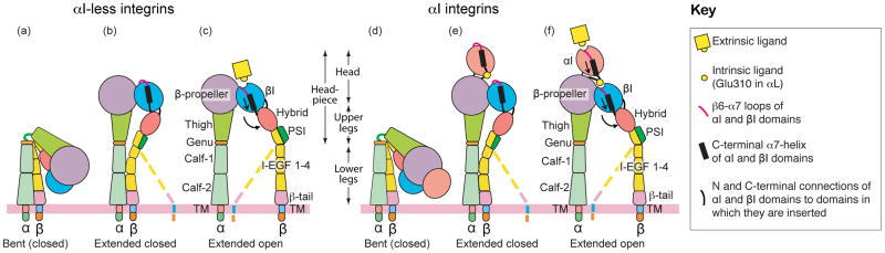
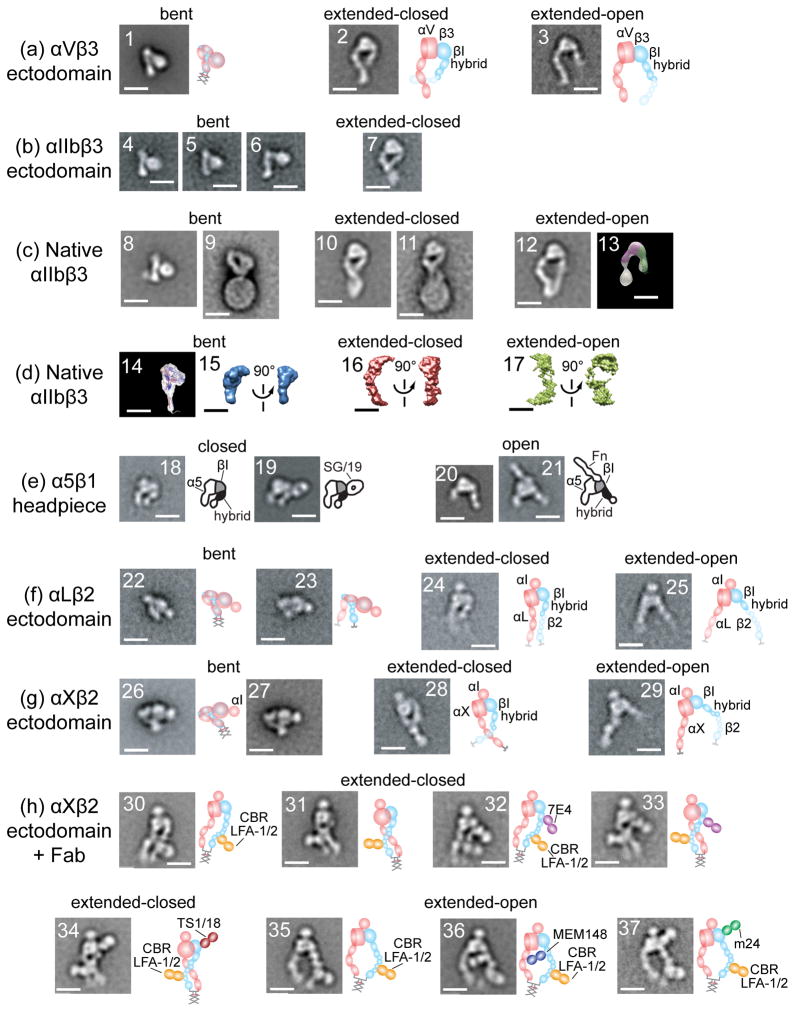
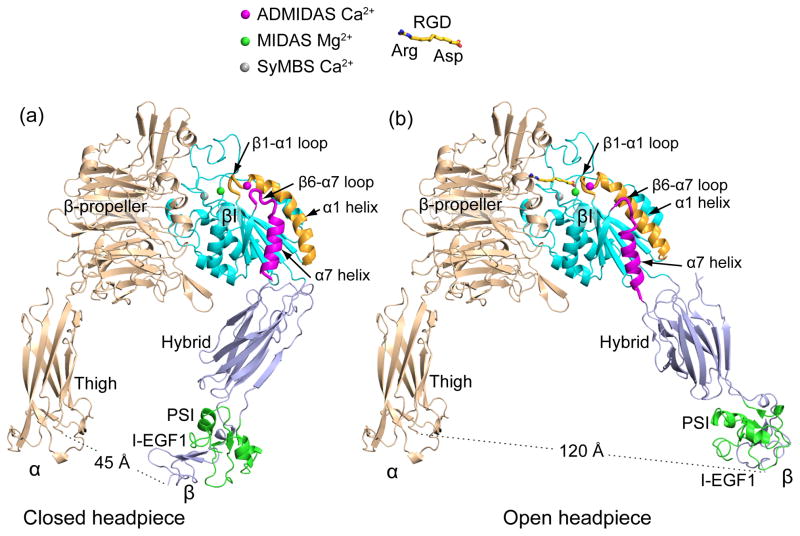
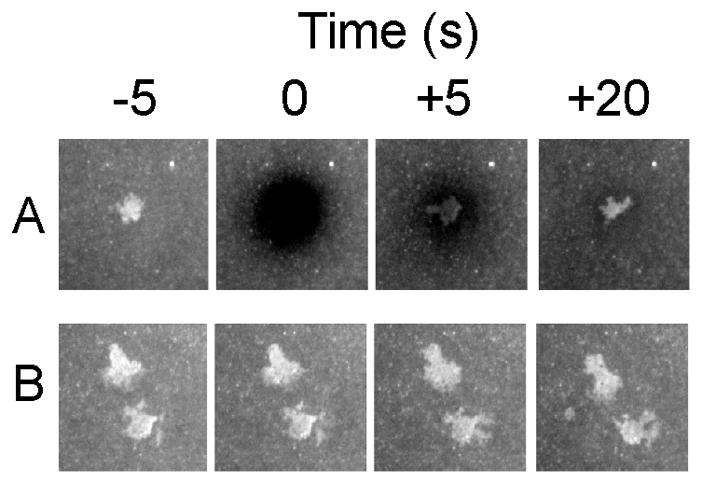
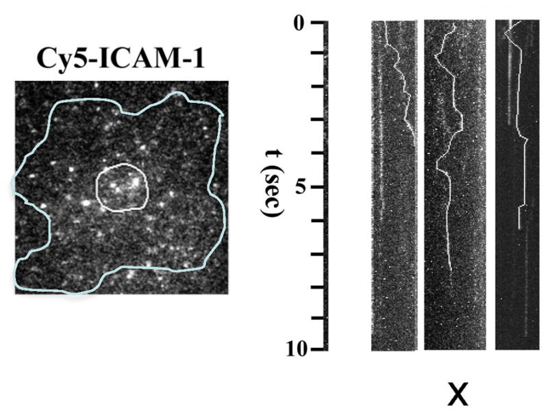
Integrins dynamically equilibrate between three conformational states on cell surfaces. A bent conformation has a closed headpiece. Two extended conformations contain either a closed or an open headpiece. Headpiece opening involves hybrid domain swing-out and a 70 Å separation at the integrin knees, which is conveyed by allostery from the hybrid-proximal end of the βI domain to a 3 Å rearrangement of the ligand-binding site at the opposite end of the βI domain. Both bent-closed and extended-closed integrins have low affinity, whereas extended-open integrin affinity is 10(3) to 10(4) higher. Integrin-mediated adhesion requires the extended-open conformation, which in physiological contexts is stabilized by post-ligand binding events. Integrins thus discriminate between substrate-bound and soluble ligands. Analysis of LFA-1-ICAM-1 interactions in the immunological synapse suggests that bond lifetimes are on the order of seconds, which is consistent with high affinity interactions subjected to cytoskeletal forces that increase the dissociation rate. LFA-1 βI domain antagonists abrogate function in the immunological synapse, further supporting a critical role for high affinity LFA-1.


Figures

Figure 1
Figure 1
The three overall integrin conformational states. The bent conformation has a closed headpiece and is low affinity. Extension at the α- and β-knees releases an interface between the headpiece and lower legs and yields an extended-closed conformation also with low affinity. Swing-out of the hybrid domain at its interface with the βI domain is connected through the βI α7-helix to rearrangements at the βI interface with the β-propeller domain that greatly (~1,000-fold) increase affinity for ligand in the extended-open conformation. Similar interdomain rearrangements in αI integrins result in activating a binding site for an internal ligand, Glu310 in αL, which pulls down the αI α7-helix to activate a similar increase in affinity (~1,000 to 10,000-fold) of the αL I domain for the ligand ICAM-1. Although the integrin headpiece has highly preferred closed and open conformations, the lower β-legs are highly flexible, and thus we speak of “overall” conformational states. This is symbolized by the dashed lower β-leg. Therefore, only very large separations between α and β TMD, such as induced by lateral motion of β when its cytoplasmic domain is associated with the actin cytoskeleton, can be transmitted through the floppy β-leg to stabilize the high-affinity, open headpiece conformation.
Figure 2
Figure 2
Structure of integrins from electron microscopy, electron tomography, and neutron or X-ray scattering in solution reveal three conformational states. Schematics are shown to right. All scale bars = 10 nm. With permission from cited references. Panels show representative class averages of negatively-stained integrins unless otherwise noted. (a) αVβ3 ectodomain with a C-terminal coiled-coil clasp (1) or unclasped with Mn2+ (2) or RGD (3) [13]. (b) αIIbβ3 ectodomain, clasped (4) or unclasped (5-7) [3]. (c) αIIbβ3 purified from platelets in detergent (8, 10, 12) [23], (13) [16] and embedded in lipoprotein nanodiscs (9, 11) [27]. With no additions (8-9), Mn2+ (10), talin head domain (11), and RGD peptide or RGD mimetics (12-13). Negative stain electron tomography class average (13). (d) Three-dimensional molecular envelopes of purified, detergent-soluble native αIIbβ3 in solution determined by small-angle neutron scattering (14) [21] or X-ray scattering (15-17) [23] with no additions (14-15), Mn2+ (16), or Mn2+ and RGD mimetic (17). (e) α5β1 headpiece [14,15] alone (18), + allosteric inhibitory SG/19 Fab (19), + RGD (20), or + fibronectin domain 7-10 fragment (21). (f) LFA-1 ectodomain [19] clasped (22) or unclasped (23-25). (g) αXβ2 [17,19] clasped from different publications (26-27) and unclasped (28-29). (h) αXβ2 [17,19] clasped with extension and activation-promoting CBR LFA-1/2 Fab (30, 31, 35); and with CBR LFA-1/2 Fab and allosteric inhibitory Fab 7E4 (32-33) and TS1/18 (34) or allosteric activating Fab MEM148 (36) or m24 (37).
Figure 3
Figure 3
The closed and open integrin headpiece conformations. A 2.3 Å movement of the βI-αI loop at the ligand binding site that raises affinity for ligand 1,000 to 10,000-fold is amplified to 4 Å displacement of the ADMIDAS Ca2+, 5 Å sideways displacement of the αI-helix as it straightens, 7 Å reshaping of the β6-α7 loop, 5.5 Å connecting rod-like displacement of the α-7 helix, and leverages hybrid domain swing-out to a 75 Å separation at the integrin knees. Structures are of the αIIbβ3 headpiece crystallized in absence [12] (a) or presence [11] (b) of RGD peptide with missing portions supplemented by superposition of the αIIbβ3 ectodomain [3].
Figure 4
Figure 4
Dynamics of LFA-1-ICAM-1 interactions. Naïve T cells adhered to supported planar bilayers with 200 molecules/μm2 Cy5-ICAM-1 and 20 molecules/μm2 agonist MHC-peptide complexes. A. At T = 0 the ICAM-1 fluorescence was bleached and the contact area was imaged at +5 and +20 seconds. B. Control without bleaching. Courtesy of T. N. Sims.
Figure 5
Figure 5
Imaging single ICAM-1 molecules in the synapse. Single Cy5-ICAM-1 imaged in an immune synapse (outline) with ~0.1% labeling. Kymographs for single particles show time vs. x-component of random x-y movement and periods of binding (vertical segments). Courtesy of R. Varma.

Similar articles

Cited by

References

    1. Xiong J-P, Stehle T, Diefenbach B, Zhang R, Dunker R, Scott DL, Joachimiak A, Goodman SL, Arnaout MA. Crystal structure of the extracellular segment of integrin αVβ3. Science. 2001;294:339–345. - PMC - PubMed
    1. Xiong JP, Mahalingham B, Alonso JL, Borrelli LA, Rui X, Anand S, Hyman BT, Rysiok T, Muller-Pompalla D, Goodman SL, et al. Crystal structure of the complete integrin αVβ3 ectodomain plus an α/β transmembrane fragment. J Cell Biol. 2009;186:589–600. - PMC - PubMed
    1. Zhu J, Luo BH, Xiao T, Zhang C, Nishida N, Springer TA. Structure of a complete integrin ectodomain in a physiologic resting state and activation and deactivation by applied Forces. Mol Cell. 2008;32:849–861. - PMC - PubMed
    1. Xie C, Zhu J, Chen X, Mi L, Nishida N, Springer TA. Structure of an integrin with an αI domain, complement receptor type 4. EMBO J. 2010;29:666–679. - PMC - PubMed
    1. Luo B-H, Springer TA, Takagi J. High affinity ligand binding by integrins does not involve head separation. J Biol Chem. 2003;278:17185–17189. - PubMed