Review
. 2011 Dec 1;10(12):945-63.
doi: 10.1038/nrd3599.

Taking aim at the extracellular matrix: CCN proteins as emerging therapeutic targets

Affiliations
Review

Taking aim at the extracellular matrix: CCN proteins as emerging therapeutic targets

Joon-Il Jun et al. Nat Rev Drug Discov. .

Abstract

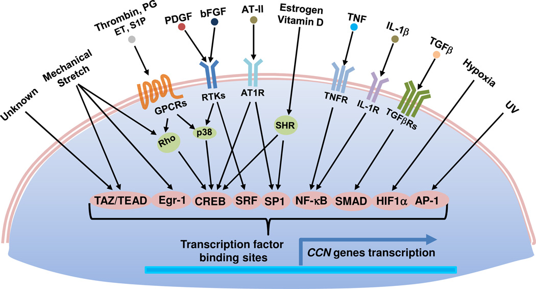
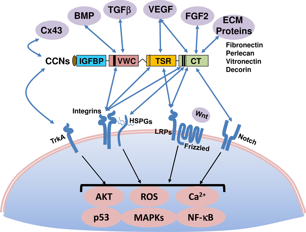
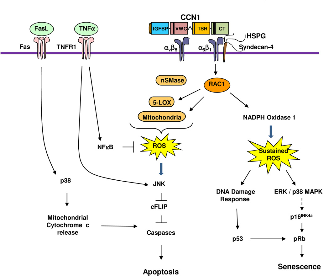
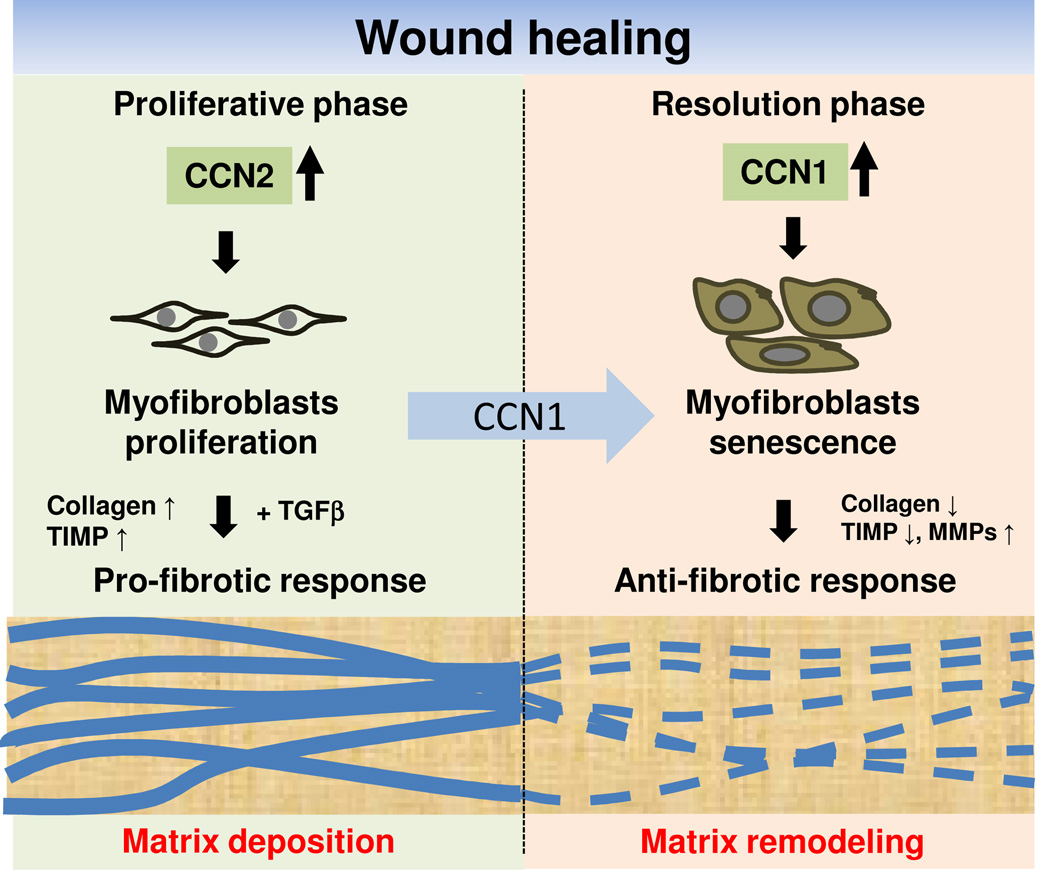
Members of the CCN family of matricellular proteins are crucial for embryonic development and have important roles in inflammation, wound healing and injury repair in adulthood. Deregulation of CCN protein expression or activities contributes to the pathobiology of various diseases - many of which may arise when inflammation or tissue injury becomes chronic - including fibrosis, atherosclerosis, arthritis and cancer, as well as diabetic nephropathy and retinopathy. Emerging studies indicate that targeting CCN protein expression or signalling pathways holds promise in the development of diagnostics and therapeutics for such diseases. This Review summarizes the biology of CCN proteins, their roles in various pathologies and their potential as therapeutic targets.


Figures

Figure 1
Figure 1. Transcriptional regulation of CCN genes
Multiple extracellular and environmental stimuli rapidly induce CCN genes through activation of various transcription factors, usually without requiring de novo protein synthesis. These include PDGF, FGF2, TGF-β, IL1β, TNFα, angiotensin II (AT-II), , agonists of G protein-coupled receptors (GPCRs) such as thrombin, prostaglandins (PE), endothelin-1 (ET), and sphingosine-1-phosphate (S1P), hormones (estrogen and vitamin D) that which bind steroid hormone receptors (SHR), hypoxia, UV, and mechanical stretch, . The locations of various transcription factor bindings sites vary for each CCN gene.
Figure 2
Figure 2. Molecular interactions through modular domains of CCN proteins
CCNs physically interact with a number of ECM proteins (including fibronectin, perlecan, vitronectin, decorin), growth factors (including such as VEGF, FGF2, TGF-β, and BMPs), and the gap junction protein connexin43, . The specific modular domains mediating the interactions, where elucidated, are indicated. Whereas fibronectin and perlecan bind the CT domain, whether decorin and vitronectin also bind CT is not as clear. CCNs also bind to and signal through a number of cell surface receptors including several integrins, , which function in concert with HSPGs or LRPs as coreceptors in some contexts. CCN2 also binds TrkA in a complex with β1 integrins as co-receptors, and CCN3 can bind Notch. CCNs can modulate Wnt signaling, in part through binding to the Wnt coreceptor, LRP-6. The activities of the CCN modular domains may interact in a combinatorial manner to induce unique activities and functions. For example, CCN1 synergism with TNFα requires its binding to both αvβ5 and α6β1 integrins through the VWC and CT domains, respectively, to induce signals from both integrins that converge within the cell; activation of either integrin alone is insufficient.
Figure 3
Figure 3. Signaling mechanism of CCN1-induced senescence and crosstalk with TNFα and FasL
The binding of CCN1 to integrin α6β1-HSPGs (syndecan 4) triggers the activation of RAC-1 and NADPH oxidase 1, leading to a much more robust and sustained level of ROS compared to cell adhesion to other ECM proteins. The sustained ROS induces a DNA damage response and the activation of p53, and triggers the activation of ERK and p38 MAPK, which in turn induces p16INK4a and activates pRb. Activated p53 and pRb contribute to the induction of cellular senescence. If integrin αvβ5 is also engaged by CCN1, RAC1-dependent ROS accumulation includes contribution from 5-lipoxygenase (5-LOX) and the mitochondria; neutral sphingomyelinase (nSMase) also contributes to CCN1-induced ROS. CCN1-induced ROS counteracts the effect of NFκB, which is strongly activated by TNFα and induces the expression of antioxidant proteins. The high level of ROS inhibits cysteine phosphatases that can inactivate MAPKs such as JNK, ERK, and p38 MAPK, leading to a hyperactivation of these kinases. Activated JNK targets the proteosomal degradation of cFLIP, an inhibitor of caspase activation, allowing the activation of caspases 8/10 by TNFα to induce apoptosis without blocking de novo protein synthesis or NFκB signaling. In addition to CCN1, CCN2 and CCN3 also synergize with TNFα to induce apoptosis, presumably through a similar mechanism. In the presence of FasL, which can trigger apoptosis on its own, CCN1 or CCN2-induced ROS leads to the hyperactivation of p38 MAPK, which enhances cytochrome c release from the mitochondria and thereby increases apoptosis.
Figure 4
Figure 4. Role of CCNs in wound healing
CCN1 and CCN2 have distinct roles in wound healing. CCN2 functions in the granulation tissue during the proliferative phase and acts with TGF-β to promote the synthesis ECM, leading to a pro-fibrotic response. As wound healing progresses, CCN1 accumulates to a sufficiently high level to induce an anti-fibrotic senescence switch in myofibroblasts, thereby limiting fibrosis by converting the ECM-synthesizing myofibroblasts into ECM-degrading senescent cells.

References

    1. Bornstein P, Sage EH. Matricellular proteins: extracellular modulators of cell function. Curr. Opin. Cell Biol. 2002;14:608–616. - PubMed
    1. Chen C-C, Lau LF. Functions and Mechanisms of Action of CCN Matricellular Proteins. Int. J. Biochem. Cell Biol. 2009;41:771–783. - PMC - PubMed
    1. Leask A, Abraham DJ. All in the CCN family: essential matricellular signaling modulators emerge from the bunker. J. Cell Sci. 2006;119:4803–4810. - PubMed
    1. Holbourn KP, Acharya KR, Perbal B. The CCN family of proteins: structure-function relationships. Trends Biochem. Sci. 2008;33:461–473. - PMC - PubMed
    1. Perbal B, Takigawa M. CCN Proteins: A New Family of Cell Growth and Differentiaion Regulators. London: Imperial College Press; 2005.

Publication types

MeSH terms

Substances