. 2011 Dec 1;2(12):e235.
doi: 10.1038/cddis.2011.116.

Novel tumor suppressive function of Smad4 in serum starvation-induced cell death through PAK1-PUMA pathway

Affiliations

Novel tumor suppressive function of Smad4 in serum starvation-induced cell death through PAK1-PUMA pathway

S-H Lee et al. Cell Death Dis. .

Abstract

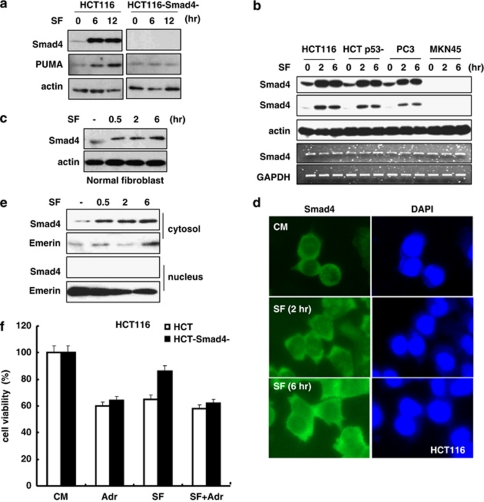
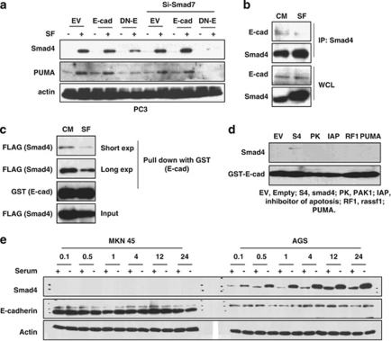
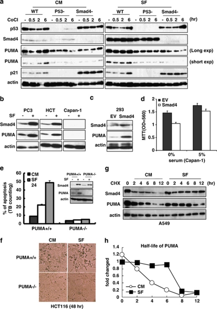
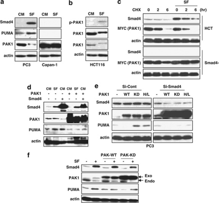
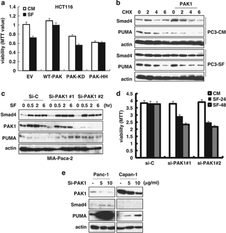
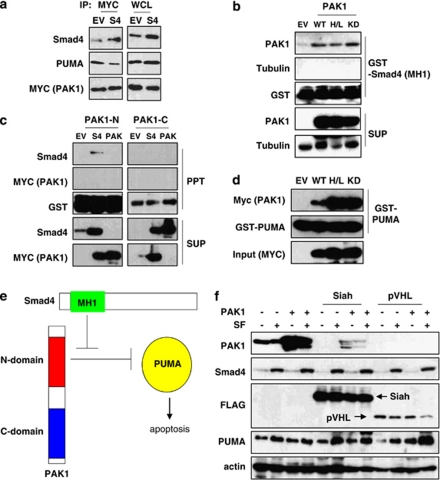
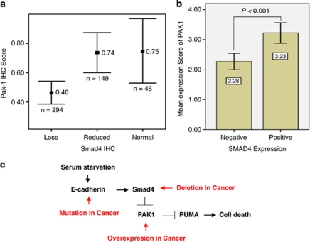
DPC4 (deleted in pancreatic cancer 4)/Smad4 is an essential factor in transforming growth factor (TGF)-β signaling and is also known as a frequently mutated tumor suppressor gene in human pancreatic and colon cancer. However, considering the fact that TGF-β can contribute to cancer progression through transcriptional target genes, such as Snail, MMPs, and epithelial-mesenchymal transition (EMT)-related genes, loss of Smad4 in human cancer would be required for obtaining the TGF-β signaling-independent advantage, which should be essential for cancer cell survival. Here, we provide the evidences about novel role of Smad4, serum-deprivation-induced apoptosis. Elimination of serum can obviously increase the Smad4 expression and induces the cell death by p53-independent PUMA induction. Instead, Smad4-deficient cells show the resistance to serum starvation. Induced Smad4 suppresses the PAK1, which promotes the PUMA destabilization. We also found that Siah-1 and pVHL are involved in PAK1 destabilization and PUMA stabilization. In fact, Smad4-expressed cancer tissues not only show the elevated expression of PAK1, but also support our hypothesis that Smad4 induces PUMA-mediated cell death through PAK1 suppression. Our results strongly suggest that loss of Smad4 renders the resistance to serum-deprivation-induced cell death, which is the TGF-β-independent tumor suppressive role of Smad4.


Figures

Figure 1
Figure 1
Induction of Smad4 in response to serum starvation. (a) Induction of Smad4 and PUMA in response to SF condition. HCT116 and its isogenic Smad4-deficient cells were incubated in SF condition for indicating time. Actin was used as loading control. (b) Serum starvation leads to Smad4 induction in three kinds of cell lines, but not in MKN45, at post-transcription level. Smad4 expression was determined by WB analysis at translation level and by reverse transcription (RT)-PCR analysis at transcription level. (c) Induction of Smad4 is observed in normal fibroblast. Normal human fibroblast was exposed to SF condition for indicating time, and the Smad4 expression level was evaluated by WB. (d) Serum starvation-induced Smad4 is accumulated in cytosol. After fixation with Me-OH, cells were stained with anti-Smad4 (green) and 4′6-diamidino-2-phenylindole (DAPI; blue). (e) Smad4 is retained in cytosol. HCT116 cells were divided into four subcellular fractions using a Sub cell kit (Calbiochem), and the expressions of Smad4 were examined. (f) Smad4-deficient HCT116 cells showed resistance to SF condition compared with positive cells, but in the presence of DNA-damaging agent, adriamycin (Adr; 2 μg/ml), both cells revealed similar sensitivity. Cell viability was determined by MTT assay
Figure 2
Figure 2
E-cad is involved in Smad4 regulation. (a) Blocking the E-cad reduces the Smad4 induction in response to SF condition. PC3 cells were transfected with empty vector (EV), E-cad, DN-E-cad (DN-E), and siRNA Smad7. (b) Interaction between Smad4 and E-cad was decreased in SF condition. At 24 h after transfection, HEK293 cell lysates were immunoprecipitated with anti-Smad4 and analyzed by WB. (c) Smad4 is released from E-cad in SF condition. GST–E-cad was incubated with Smad4–FLAG-transfected HEK293 cell lysates in RIPA buffer for 2 h. After washing, precipitated protein was subjected to SDS-PAGE and WB with the indicated antibodies. (d) Interaction between Smad4 and E-cad is a specific event. GST pull down-was performed with several proteins, and each protein was analyzed by detectable antibodies (data not shown). (e) In the serum-deficient condition, Smad4 induction is only shown in AGS cells (containing wild-type E-cad), but not in mutant E-cad harboring MKN45. Each cell line was exposed to SF condition for indicated times, and Smad4 and E-cad proteins were detected by WB
Figure 3
Figure 3
Smad4 induces PUMA. (a) PUMA is induced by Smad4 in response to SF condition. PUMA expression level is significantly decreased not only in p53-deficient cells but also in Smad4-deficient cells. Expression of indicated proteins in HCT116 cells was assessed by WB. (b) Smad4-dependent induction of PUMA. PUMA induction was not detected in Smad4-deficient Capan-1 cells. (c) Overexpression of Smad4 induces PUMA expression. HEK293 cells were transfected with empty vector (EV) and Smad4, and indicating proteins were detected by WB analysis. (d) Smad4 transfection can restore the SF-induced cell death in Smad4-deficient Capan-1. Cells were transfected with EV and Smad4 for 24 h, and viability in different serum concentration was determined by MTT assay. (e and f) Absence of apoptosis in PUMA-deficient HCT116. HCT116 (PUMA+/+) and its isogenic PUMA-deficient cells (PUMA−/−) were incubated in SF condition for 24 or 48 h (f). Dying cells were counted using trypan blue dye exclusion assay. PUMA deficiency did not alter the Smad4 induction by SF condition (inset). (g) Extension of protein half-life of Smad4 and PUMA in SF condition. A549 cells were incubated in SF condition for 6 h to adjust the starting protein levels and were incubated in SF or complete media condition for indicated time with CHX (100 μg/ml). (h) The half-life of the PUMA protein is remarkably increased in SF condition. Expression level of PUMA was determined by densitometer. Presented graph showed the fold changes compared with control (CHX 0 time in SF condition)
Figure 4
Figure 4
Smad4 regulates PUMA through PAK1. (a) PAK1 is reduced in SF condition. Cells were incubated with SF condition for 6 h. (b) In response to serum starvation, phoshorylated PAK1 (p-PAK1) is increased, whereas total PAK1 is reduced. HCT116 cells were incubated with SF condition for 6 h. (c) PAK1 expression is suppressed by Smad4. Compared with Smad4-deficient HCT116 cells, Smad4 stability was increased, whereas PAK1 expression was reduced in response to SF. CHX was treated with each cell line, and Smad4 and PAK1 (Myc tagged) expression levels were examined by WB analysis. (d) Smad4 overexpression induces PUMA via suppression of PAK1 expression. However, elevated PAK1 expression could attenuate Smad4-mediated PUMA induction in SF condition. HEK293 was transfected with wild-type PAK1 and Smad4 for 24 h. (e) Knockdown of Smad4 using siRNA-induced PAK1 expression. siRNA Smad4, wild-type PAK1, kinase-dead PAK1 (KD-PAK1; K299R), and H/L (H83LH86L; GTPase binding domain mutant) was transfected to PC3 for 24 h, and indicated proteins were measured by WB. (f) PAK1 mutants could not suppress Smad4-mediated PUMA induction in SF condition. Expression of PUMA was repressed by wild-type PAK1 but not by KD-PAK1
Figure 5
Figure 5
PAK1 regulates SF-induced cell death and PUMA expression. (a) Serum deprivation-induced cell death is reduced by wild-type PAK1, but not by mutant PAK1. Cell viability in SF condition was checked by MTT assay in HCT116 after transfection with PAK1s. (b) The stability (half-life) of PUMA is decreased by overexpression of wild-type PAK1. PC3 cells were transfected with wild-type PAK1, and cell lysates were analyzed by WB after incubating with CHX. (c) PAK1 knockdown restores PUMA induction in response to SF condition. MIA-Paca-2 cells were transfected with si-PAK1 for 24 h, and expressions of indicated proteins were evaluated by WB analysis. (d) Elimination of PAK1 increases the sensitivity to serum starvation-induced cell death. siRNA PAK1 no. 1 and no. 2 were transfected for 24 h before exposing to SF condition. Cell viability was determined by MTT assay. (e) PAK1 knockdown induces PUMA expression in pancreatic cancer cell lines. Cells were transfected with a range of si-PAK1 concentration for 24 h, and PUMA induction by disruption of PAK1 is revealed even in Smad4-null cell, Capan-1. Expression of indicating proteins were assessed by WB
Figure 6
Figure 6
Direct interaction between PUMA and PAK1–Smad4. (a) PAK1 interacts with Smad4 and PUMA. HEK293 lysate overexpressing wild-type PAK1 (Myc tagged) was used for IP, which was performed with anti-Myc. PAK1 can bind to both Smad4 and PUMA, but overexpression of Smad4 attenuates PAK1 and PUMA interaction. (b) PAK1 binds to Smad4 MH1 domain. Agarose bead-conjugated GST–Smad4 (MH1) was incubated with PAK1-transfected HEK293 lysates, and Smad4-associated PAK1 was determined by WB after precipitation. Tubulin was used as a negative control. (c) N terminus of PAK1 is essential to bind to Smad4. GST pull-down was performed with PAK1 N- and C- terminal fragments. (d) Both wild and mutant PAK1 can bind to PUMA. PAK1 (Myc tagged) expressing HEK293 lysates were used for GST pull-down. (e) Interaction map between Smad4 and PAK1 and PUMA–PAK1. MH1 domain of Smad4 bound to and protected the PAK1-N-terminal domain-induced PUMA suppression. (f) Siah and pVHL reduced PAK1 but induced PUMA expression. Siah (FLAG tagged) and pVHL (FLAG tagged) were transfected to HEK293 for 24 h before eliminating serum from media. Expression of indicating proteins were examined by WB analysis
Figure 7
Figure 7
Elevated expression of PAK1 in Smad4-positive colon cancer tissues. (a) Analysis of PAK1 expression in 489 human colon cancer tissues using IHC analysis. In Smad4-intact tissues, PAK1 expression was elevated. (b) PAK1 expression is elevated in Smad4-negative tissues. More detail clinicopathological data were available in Supplementary Table 2. (c) Diagram for summary. In response to serum deprivation, Smad4 is induced by releasing from E-cad and suppresses PAK1-mediated PUMA reduction, which causes induction and activation of apoptosis program. Against Smad4-mediated cell death, cancer cells evoke the mutation or deletion of Smad4 itself, or mutation of E-cad, or overexpression of PAK1. Overcome mechanisms of smad4-induced cell death are depicted as red letters

Similar articles

Cited by

References

    1. Weinberg RA. How cancer arises. Sci Am. 1996;275:62–70. - PubMed
    1. Vogelstein B, Kinzler KW. Cancer genes and the pathways they control. Nat Med. 2004;10:789–799. - PubMed
    1. Bardeesy N, DePinho RA. Pancreatic cancer biology and genetics. Nature Rev Cancer. 2002;2:897–909. - PubMed
    1. Lei M, Lu W, Meng W, Parrini MC, Eck MJ, Mayer BJ, et al. Structure of PAK1 in an autoinhibited conformation reveals a multistage activation switch. Cell. 2000;102:387–397. - PubMed
    1. Hahn SA, Schutte M, Hoque AT, Moskaluk CA, da Costa LT, Rozenblum E, et al. DPC4, a candidate tumor suppressor gene at human chromosome 18q21.1. Science. 1996;271:350–353. - PubMed

Publication types

MeSH terms