Review
. 2012 Feb;22(2):116-24.
doi: 10.1016/j.tcb.2011.10.003. Epub 2011 Nov 28.

A PLA1-2 punch regulates the Golgi complex

Affiliations
Review

A PLA1-2 punch regulates the Golgi complex

Marie E Bechler et al. Trends Cell Biol. 2012 Feb.

Abstract

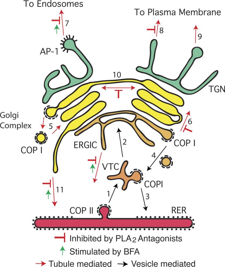
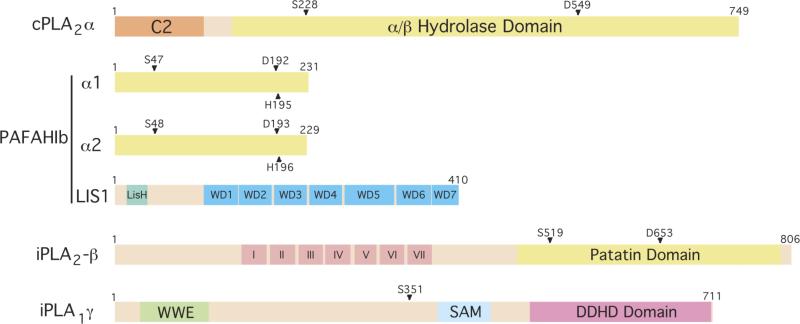
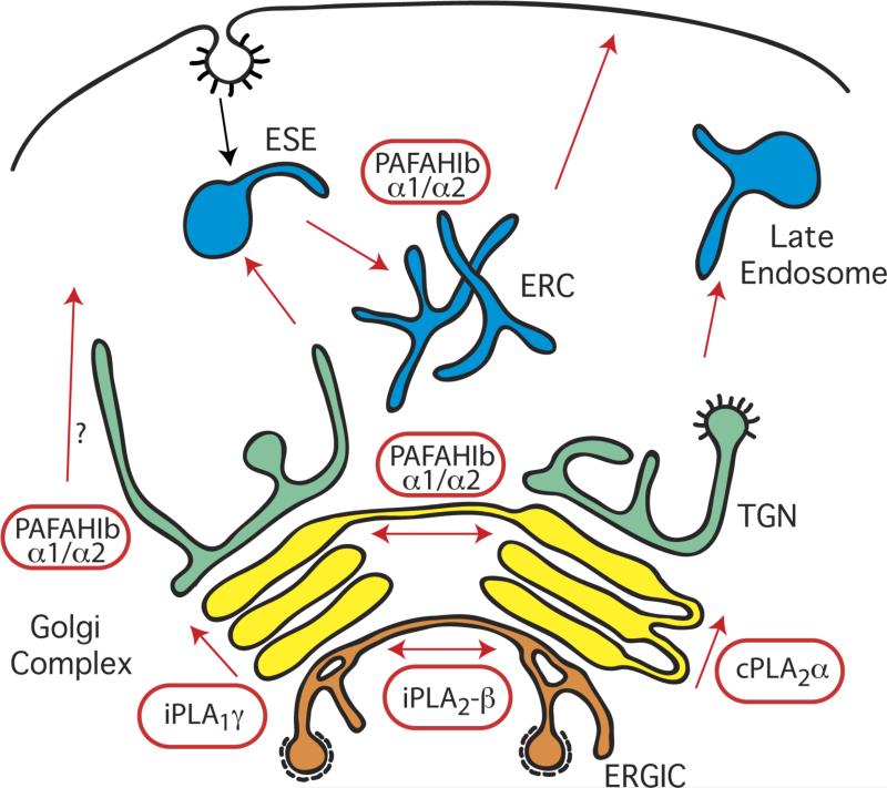
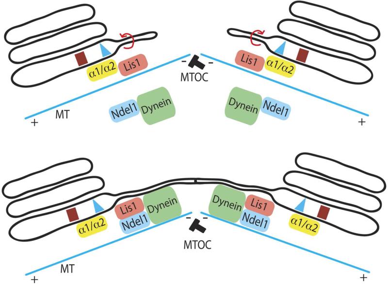
The mammalian Golgi complex, trans Golgi network (TGN) and ER-Golgi intermediate compartment (ERGIC) are comprised of membrane cisternae, coated vesicles and membrane tubules, all of which contribute to membrane trafficking and maintenance of their unique architectures. Recently, a new cast of players was discovered to regulate the Golgi and ERGIC: four unrelated cytoplasmic phospholipase A (PLA) enzymes, cPLA(2)α (GIVA cPLA(2)), PAFAH Ib (GVIII PLA(2)), iPLA(2)-β (GVIA-2 iPLA(2)) and iPLA(1)γ. These ubiquitously expressed enzymes regulate membrane trafficking from specific Golgi subcompartments, although there is evidence for some functional redundancy between PAFAH Ib and cPLA(2)α. Three of these enzymes, PAFAH Ib, cPLA(2)α and iPLA(2)-β, exert effects on Golgi structure and function by inducing the formation of membrane tubules. We review our current understanding of how PLA enzymes regulate Golgi and ERGIC morphology and function.


Figures

Figure I
Figure I
A simplified diagram of the Golgi complex as a hub of membrane trafficking.
Figure 1
Figure 1. Domain structures of Golgi- and ERGIC-associated PLA enzymes
cPLA2α contains a large α/β hydrolase domain with residues S228 and D549 comprising the active site dyad. It also contains a C2 Ca2+-binding domain that is necessary for translocation to Golgi membranes. PAFAH Ib is comprised of α1 and α2 homo- or heterodimers together with LIS1. The indicated residues form the active site triad. iPLA2–β contains a patatin lipase domain, with an active site at S519, and seven ankyrin repeats (I-VII). iPLA1γ contains an S351 residue, which when mutated to alanine (A) abolishes catalytic activity, a DDHD2 domain that is conserved with iPLA1β, a WWE domain predicted to mediate protein-protein interactions in ubiquitination and ADP-ribosylation systems, and a SAM domain (sterile alpha motif) that can mediate both homo- and hetero-oligomerization.
Figure 2
Figure 2. PLA enzymes that regulate the structure and function of the Golgi complex and endosomes
cPLA2α localizes to the Golgi complex and mediates the formation of membrane tubules that facilitate anterograde transport through the cisternal stack. The α1 and α2 PLA2 catalytic subunits of PAFAH Ib localize to multiple Golgi cisternae, early sorting endosomes (ESEs), and the endocytic recycling compartment (ERC). These enzymes contribute to membrane tubule formation that links cisternal stacks into intact Golgi ribbons and to those that facilitate endocytic recycling of transferrin and its receptor from ESEs and the ERC. In addition, loss of α1 and α2 inhibits export from the TGN, although it is not clear if this involves membrane tubules. iPLA2–β localizes specifically to the ERGIC where it mediates the formation of membrane tubules that bridge between separate ERGIC clusters. iPLA1γ appears to localize primarily to the cis Golgi and may influence anterograde transport through the cisternal stack.
Figure 3
Figure 3. Model integrating membrane curvature produced by the PLA2 activity of PAFAH Ib α1 and α2 with Lis1-mediated dynein transport along microtubules
PAFAHIB initiates outward membrane curvature to generate a membrane tubule, which can be pulled/extended along microtubules (MT) by Lis1 and Ndel interactions with dynein. Dynein may be able to carry the membrane tubule towards the minus end of microtubules, facilitating the convergence of the Golgi stack and positioning at the microtubule organizing center (MTOC, minus end of the microtubules).

References

    1. Emr S, et al. Journeys through the Golgi--taking stock in a new era. J Cell Biol. 2009;187:449–453. - PMC - PubMed
    1. Trucco A, et al. Secretory traffic triggers the formation of tubular continuities across Golgi sub-compartments. Nat Cell Biol. 2004;6:1071–1081. - PubMed
    1. Brown WJ, et al. Phospholipase A2 (PLA2) enzymes in membrane trafficking: mediators of membrane shape and function. Traffic. 2003;4:214–221. - PubMed
    1. de Figueiredo P, et al. Evidence that phospholipase A2 activity is required for Golgi complex and trans Golgi network membrane tubulation. Proc Natl Acad Sci U S A. 1998;95:8642–8647. - PMC - PubMed
    1. de Figueiredo P, et al. Membrane tubule-mediated reassembly and maintenance of the Golgi complex is disrupted by phospholipase A2 antagonists. Mol Biol Cell. 1999;10:1763–1782. - PMC - PubMed

Publication types

Substances