

A characterization of the relationship of ovarian reserve markers with age
- PMID: 22130324
- PMCID: PMC3312467
- DOI: 10.1016/j.fertnstert.2011.10.031
A characterization of the relationship of ovarian reserve markers with age
Abstract
Objective: To identify markers of ovarian age that best match the pattern of oocyte loss seen in histology specimens.
Design: Cross-sectional study.
Setting: University.
Patient(s): Caucasian women (n = 252) aged 25-45 years.
Intervention(s): none.
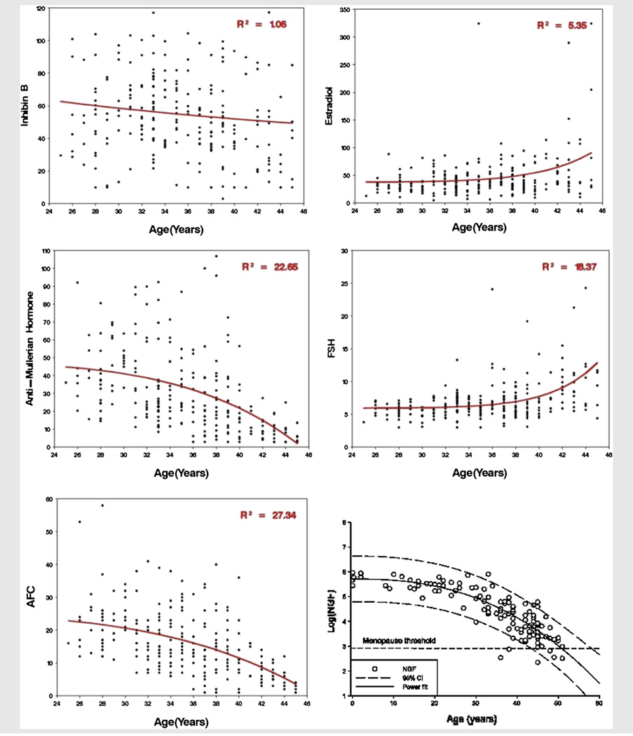
Main outcome measure(s): The relationship between antral follicle count (AFC), antimüllerian hormone (AMH), inhibin B, FSH, and E(2) with age was estimated using the power model, which previously has been shown to most accurately describe oocyte loss in histologic specimens. The power model was fit to each marker and used to compare the rates of change at ages 30 and 40 with the histologic pattern. Among those markers following the pattern, R(2) was used to compare the degree of relationship with age.
Result(s): Both AMH levels and AFC exhibited significant progressive declines with age. The average rates of loss per year for AFC and AMH were, respectively, -0.57 and -1.09 at age 30, and -1.33 and -3.06 at age 40. FSH, inhibin B, and E(2) did not exhibit progressive rates of change. The R(2) for AFC was 27.3% and for AMH was 22.7%.
Conclusion(s): Only AFC and AMH follow the pattern of oocyte loss observed histologically. Although AMH may be more cost-effective, AFC is a slightly more accurate noninvasive measure for ovarian aging.
Copyright © 2012 American Society for Reproductive Medicine. Published by Elsevier Inc. All rights reserved.
Figures
References
-
- te Velde ER, Pearson PL. The variability of female reproductive ageing. Hum Reprod Update. 2002;8:141–154. - PubMed
-
- Richardson SJ, Senikas V, Nelson JF. Follicular depletion during the menopausal transition: evidence for accelerated loss and ultimate exhaustion. J Clin Endocrinol Metab. 1987;65:1231–1237. - PubMed
-
- Faddy MJ, Gosden RG, Gougeon A, Richardson SJ, Nelson JF. Accelerated disappearance of ovarian follicles in mid-life: implications for forecasting menopause. Hum Reprod. 1992;7:1342–1346. - PubMed
-
- Block E. Quantitative morphological investigations of the follicular system in women; variations at different ages. Acta Anat (Basel) 1952;14:108–123. - PubMed
-
- Faddy MJ, Gosden RG. A model conforming the decline in follicle numbers to the age of menopause in women. Hum Reprod. 1996;11:1484–1486. - PubMed
Publication types
MeSH terms
Substances
Grants and funding
LinkOut - more resources
Full Text Sources
Medical
