. 2012 Apr;69(8):1377-89.
doi: 10.1007/s00018-011-0884-2. Epub 2011 Dec 1.

Tbx2 and Tbx3 induce atrioventricular myocardial development and endocardial cushion formation

Affiliations

Tbx2 and Tbx3 induce atrioventricular myocardial development and endocardial cushion formation

Reena Singh et al. Cell Mol Life Sci. 2012 Apr.

Abstract

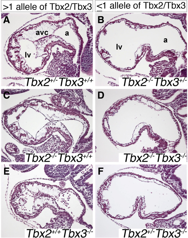
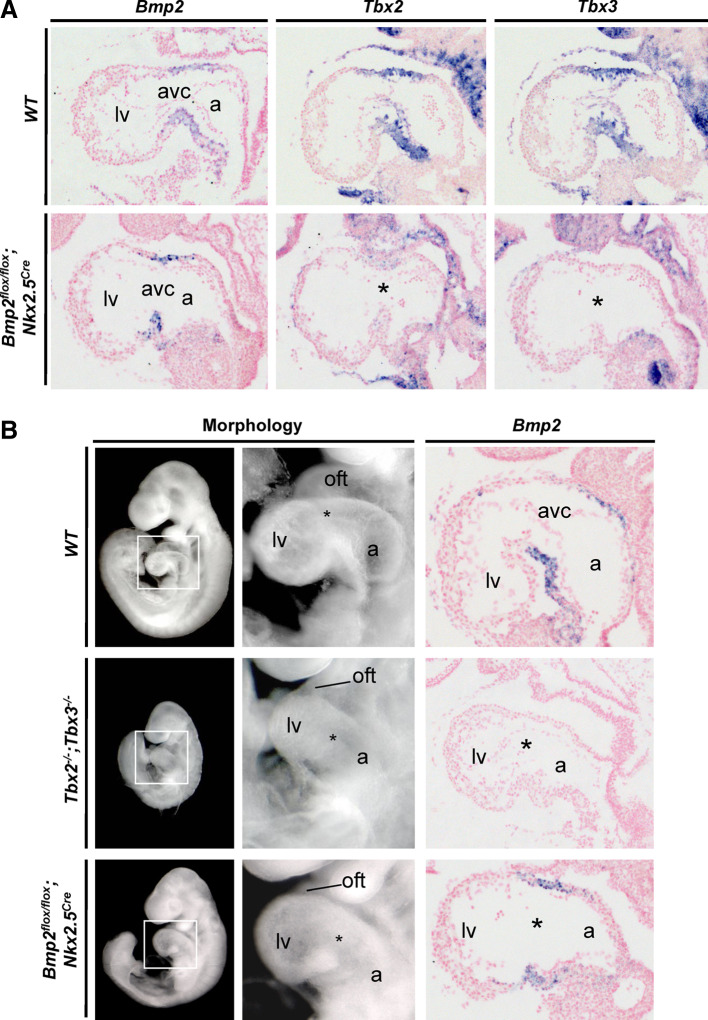
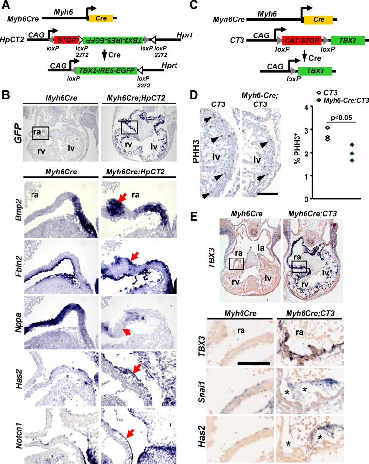
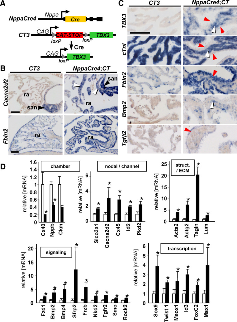
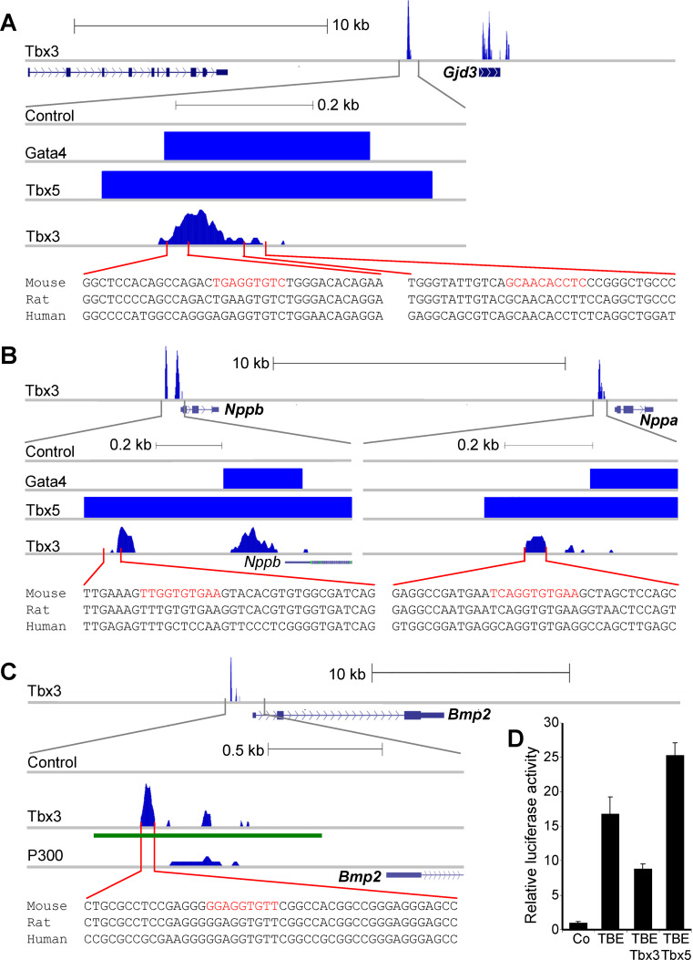
A key step in heart development is the coordinated development of the atrioventricular canal (AVC), the constriction between the atria and ventricles that electrically and physically separates the chambers, and the development of the atrioventricular valves that ensure unidirectional blood flow. Using knock-out and inducible overexpression mouse models, we provide evidence that the developmentally important T-box factors Tbx2 and Tbx3, in a functionally redundant manner, maintain the AVC myocardium phenotype during the process of chamber differentiation. Expression profiling and ChIP-sequencing analysis of Tbx3 revealed that it directly interacts with and represses chamber myocardial genes, and induces the atrioventricular pacemaker-like phenotype by activating relevant genes. Moreover, mutant mice lacking 3 or 4 functional alleles of Tbx2 and Tbx3 failed to form atrioventricular cushions, precursors of the valves and septa. Tbx2 and Tbx3 trigger development of the cushions through a regulatory feed-forward loop with Bmp2, thus providing a mechanism for the co-localization and coordination of these important processes in heart development.


Figures

Fig. 1
Fig. 1
Combined loss of Tbx2 and Tbx3 abrogates myocardial patterning of the atrioventricular canal. Comparative analysis of wild-type, Tbx2 /, Tbx2 / ;Tbx3 / and Tbx3 / embryos for cardiac morphology and molecular marker expression at E9.5. Left lateral views of whole E9.5 embryos and enlarged hearts (boxed regions in upper row shown in second row) reveal growth retardation and dilated avc phenotype in Tbx2/Tbx3 double mutant embryos. In situ hybridization analysis of marker gene expression in sagittal sections through the avc with probes as indicated. avc atrioventricular canal, la left atrium, lv left ventricle
Fig. 2
Fig. 2
Combined loss of more than two alleles of Tbx2 and Tbx3 abrogates cushion formation in the AVC. Histological analysis of sagittal sections through the left atrium (la), atrioventricular canal (avc), and left ventricle (lv) by hematoxylin and eosin (H and E) staining shows normal cushion formation upon loss of one or two functional alleles of Tbx2 and Tbx3 (a, c, e), whereas loss of more than two functional alleles of the two genes results in partial loss of cardiac jelly and complete loss of AV cushion tissue (b, d, f). avc atrioventricular canal, a atrium, lv left ventricle
Fig. 3
Fig. 3
A Bmp2-Tbx2/3 regulatory feed-forward loop for expression in the atrioventricular canal. a Inactivation of Bmp2 in cardioblasts or embryonic cardiomyocytes by Nkx25 Cre results in loss of Tbx2 and Tbx3 expression in the atrioventricular canal (asterisk). b In both compound Tbx2/Tbx3 mutants and Bmp2 conditional mutants, the atrioventricular constriction is largely absent. Bmp2 expression is lost from Tbx2/Tbx3 compound mutants, whereas reduced levels of Bmp2 transcripts (lacking exon 3; [3]) can still be found in the Bmp2 conditional mutants (asterisk). avc atrioventricular canal, a atrium, lv left ventricle
Fig. 4
Fig. 4
Cardiac misexpression of Tbx2 or Tbx3 induces cardiac jelly and cushion formation in chamber myocardium. a Use of Myh6Cre driver to ectopically activate Tbx2 in the myocardium. b Robust activation of Tbx2 (Gfp) in Myh6Cre;HpCT2 leads to loss of Nppa and induction of Bmp2 in the myocardium, and Fbln2, Has2 and Notch1 in endocardium/mesenchyme (red arrows). c Transgenic constructs used to activate Tbx3 in the myocardium of the embryonic heart. d Immunohistochemical analysis of proliferation (PHH3) in E11.5 hearts of Myh6Cre;CT3 mice compared to control (CT3) mice. Black arrows depict Phospho-H3 positive cells in the ventricular myocardium of control mice and Myh6Cre;CT3 mice. Black bar, 100 μm. e In situ hybridization of serial sections in E11.5 hearts of Myh6Cre;CT3 mice compared to control mice. Snai1 and Has2 are induced in the endocardium and subendocardial mesenchyme formed in the atria of Myh6Cre;CT3 embryos (asterisk). avc atrioventricular canal, l/ra left/right atrium, l/rv left/right ventricle
Fig. 5
Fig. 5
Myocardial Tbx3 expression induces endocardial mesenchyme formation and nodal gene expression. a Transgenic constructs used to activate TBX3 in the developing atria. b, c Sections of E17.5 atria of CT3 and NppaCre;CT3 mice were probed for expression of indicated genes. Black arrowhead indicates the sinus node (san), white arrowhead the myocardium. Red arrowheads depict the thick endocardial mesenchymal layer that forms in NppaCre;CT3 atria. Black bar, 100 μm. d qRT-PCR analysis of left atria of NppaCre;CT3 double-transgenic mice compared to CT3 control mice. Expression levels in CT3 atria were set to 1. *p < 0.05. ra right atrium, san sinus node
Fig. 6
Fig. 6
Tbx3 ChIP-seq reveals interaction with known and novel binding sites in vivo. a Tbx3 binds to the region upstream of Cx30.2 (Gjd3), a region shown to function as a T-box responsive enhancer [44]. The relative position and sequence of the published T-box binding elements and conservation are shown at the bottom of the panel (red). Recently published Gata4 and Tbx5 ChIP-seq data from HL-1 cells [46] also shows specific binding within this region. b The TBE of Nppa [42, 43], is occupied by Tbx3 in atria and by Tbx5 and Gata4 in the HL-1 cell line [46]. Tbx3, Tbx5 and Gata4 binding peaks can also be observed upstream of Nppb, a gene showing a similar expression profile and response to Tbx3. c Tbx3 binding peaks surrounding the Bmp2 gene. The upstream element, shown to bind both Tbx3 and P300 [47], was cloned (underlined in green) upstream of a minimal promoter driving luciferase. Putative T-binding sequence is shown at the bottom of the panel. d Luciferase induction in H10 cells, relative to the minimal promoter only (Co), by the putative enhancer (TBE) described in c. The activity of this element can be modulated by the presence of Tbx3 and Tbx5

References

    1. Christoffels VM, Smits GJ, Kispert A, Moorman AF. Development of the pacemaker tissues of the heart. Circ Res. 2010;106:240–254. doi: 10.1161/CIRCRESAHA.109.205419. - DOI - PubMed
    1. Combs MD, Yutzey KE. Heart valve development: regulatory networks in development and disease. Circ Res. 2009;105:408–421. doi: 10.1161/CIRCRESAHA.109.201566. - DOI - PMC - PubMed
    1. Ma L, Lu MF, Schwartz RJ, Martin JF. Bmp2 is essential for cardiac cushion epithelial–mesenchymal transition and myocardial patterning. Development. 2005;132:5601–5611. doi: 10.1242/dev.02156. - DOI - PubMed
    1. Luna-Zurita L, Prados B, Grego-Bessa J, Luxan G, Del MG, Benguria A, Adams RH, Perez-Pomares JM, de la Pompa JL. Integration of a notch-dependent mesenchymal gene program and Bmp2-driven cell invasiveness regulates murine cardiac valve formation. J Clin Invest. 2010;120:3493–3507. doi: 10.1172/JCI42666. - DOI - PMC - PubMed
    1. Walsh EP. Interventional electrophysiology in patients with congenital heart disease. Circulation. 2007;115:3224–3234. doi: 10.1161/CIRCULATIONAHA.106.655753. - DOI - PubMed

Publication types

LinkOut - more resources