. 2012 Feb;86(3):1862-73.
doi: 10.1128/JVI.06295-11. Epub 2011 Nov 30.

The insulator protein CTCF binding sites in the orf73/LANA promoter region of herpesvirus saimiri are involved in conferring episomal stability in latently infected human T cells

Affiliations

The insulator protein CTCF binding sites in the orf73/LANA promoter region of herpesvirus saimiri are involved in conferring episomal stability in latently infected human T cells

Katrin Zielke et al. J Virol. 2012 Feb.

Abstract

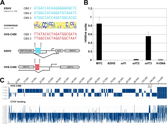
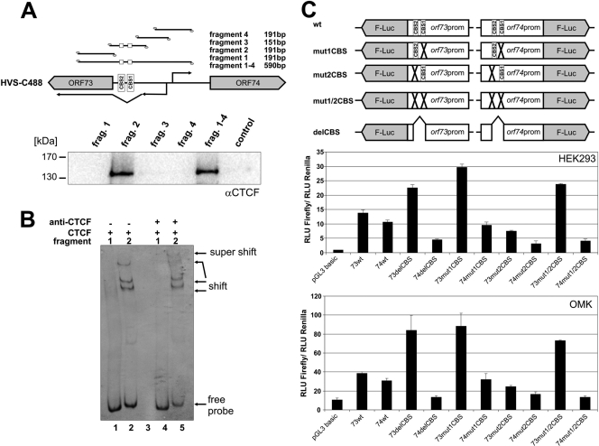
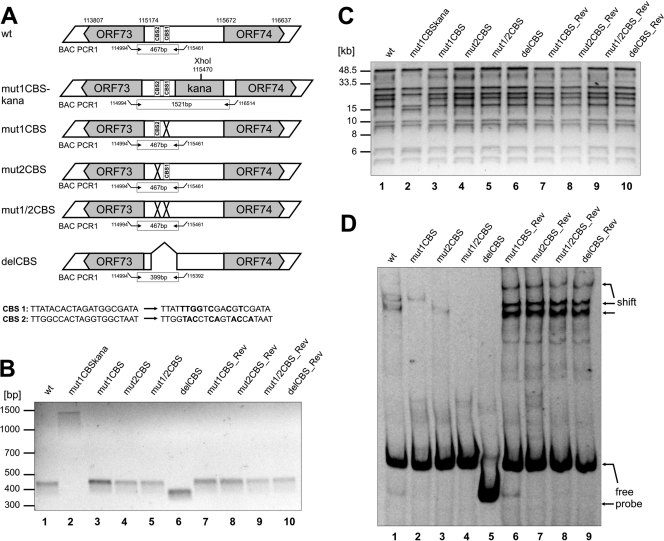
Herpesviruses establish latency in suitable cells of the host organism after a primary lytic infection. Subgroup C strains of herpesvirus saimiri (HVS), a primate gamma-2 herpesvirus, are able to transform human and other primate T lymphocytes to stable growth in vitro. The viral genomes persist as nonintegrated, circular, and histone-associated episomes in the nuclei of those latently infected T cells. Epigenetic modifications of episomes are essential to restrict the transcription during latency to selected viral genes, such as the viral oncogenes stpC/tip and the orf73/LANA. In this study, we describe a genome-wide chromatin immunoprecipitation-on-chip (ChIP-on-chip) analysis to profile the occupancy of CTCF on the latent HVS genome. We then focused on two distinct, conserved CTCF binding sites (CBS) within the orf73/LANA promoter region. Analysis of recombinant viruses harboring deletions or mutations within the CBS indicated that the lytic replication of such viruses is not substantially influenced by CTCF. However, T cells latently infected with CBS mutants were impaired in their proliferation abilities and showed a significantly reduced episomal maintenance. We detected a reduced transcription of the orf73/LANA gene in the T cells, corresponding to the reduced viral genomes; this might contribute to the loss of HVS episomes, as LANA is central in the maintenance of viral episomes in the dividing T cell populations. These data demonstrate that the episomal stability of HVS genomes in latently infected human T cells is dependent on CTCF.


Figures

Fig 1
Fig 1
Detection of putative CTCF binding sites within the intergenic region of HVS orf73/orf74. (A) Schematic comparison of published CBS within the intergenic regions of KSHV orf73/K14 (AJ410493; blue) and potential CBS at HVS orf73/orf74 (KSU756098; red) to the consensus binding sequence of CTCF (23). (B) ChIP analysis of CTCF binding to the intergenic region of HVS orf73/orf74 (exemplary analysis by B. Alberter). Viral genomes from HVS transformed human CBLs were analyzed in three independent ChIP experiments using an anti-CTCF serum (rabbit) and subsequent SYBR green PCR. Mean values were calculated for each of the three data sets, and the mean values of the single experiments were then normalized to form a combined mean value (= 1). CBS of the myc gene locus served as positive control and the first intron of the cellular ADH5 gene represents the negative control. Further coding regions of HVS (orf1 promoter region, orf72 and H-DNA) were analyzed in addition to the putative CBS at the intergenic region of orf73/orf74. (C) ChIP-on-Chip analysis of CTCF binding to the latent HVS-C488 genome using a custom oligonucleotide array (done by B. Alberter as described in reference 2). Upper panel: HVS strain C488 L-DNA containing the known 75 open reading frames and five H-DNA repeat units. The scale of the x axis corresponds to the genome position in base pairs. Lower panel: CTCF binding in the latent HVS genome. The ratio of Cy3-labeled input and Cy5-labeled ChIP probe signals along the genome are displayed using SignalMap version 1.9.
Fig 2
Fig 2
CTCF binding to the HVS orf73/orf74 promoter region influences promoter activity. (A) Oligonucleotide pulldown with biotinylated DNA from HVS C488 region 107134 to 107724. HEK293T nuclear extract was incubated with biotinylated DNA fragments and bound proteins were identified by Western blotting with an antibody specific for CTCF. (B) Fluorescent electrophoretic mobility shift assay (EMSA) with Cy5-labeled double-stranded DNA fragments encompassing fragment 1 or 2 (same fragments as in panel A), respectively. DNA fragments were incubated in the presence of recombinant CTCF protein (lanes 1 and 2) or in the presence of recombinant CTCF protein and a CTCF antibody (lanes 4 and 5) prior to gel electrophoresis. DNA fragments, fragment shifts, or supershifts were visualized by measuring the fluorescence signal. (C) Luciferase reporter assays. HEK293 cells or OMK cells were transfected with equal DNA amounts of empty vector pGL3 basic or one of the plasmids depicted in the upper scheme. A cotransfected renilla luciferase construct served as the internal standard for transfections. Luciferase assays were performed 1 day after transfection. Bars represent arithmetic means and standard deviations of luciferase activity normalized to renilla activity from three independent transfection experiments.
Fig 3
Fig 3
Generation and characterization of recombinant HVS harboring point mutations or deletions within the CBS of the intergenic region of HVS orf73/orf74. (A) Schematic diagram illustrating the structure of recombinant viruses that were generated by homologous recombination in E.coli. The upper part of panel A shows the genomic region of HVS strain C488 that contains CBS (numbers refer to nucleotide positions of HVS bacmid Bac43wt; BAC PCR1 refers to PCR amplification that was performed to confirm the integrity of recombinant viruses). The lower panels illustrate the recombinant viruses with indications of mutations/deletions of CBS (CBS 1, CTCF binding site 1; CBS 2, CTCF binding site 2; X, mutation; ⋀, deletion). For verification of the correct recombination sites within the HVS genome, all bacmid clones were analyzed by PCR and XhoI digest. (B) PCR analyses using oligonucleotides specific for the intergenic region orf73/orf74. The localization of primers used for amplification is shown in panel A (BAC PCR1). BAC PCR amplifies a fragment of 467 bp for the wild type, point mutants, and revertants or of 399 bp for the deletion mutant. Bacmids still harboring the aphAI gene display a PCR fragment of approximately 1.5 kb. The agarose gel shows the wild type (lane 1), mutant (lanes 3 to 6), and revertant (lanes 7 to 10) as well as a representative clone Bac43mut1CBS-kana (lane2) comprising the aphAI gene. (C) XhoI digest, pulse-field gel electrophoresis and subsequent ethidium bromide staining of 0.8% agarose gels of bacterial clones harboring the indicated bacmids. The aphAI cassette contains a single XhoI restriction site resulting in a different restriction pattern of bacmids containing the aphAI gene compared to the wild type, mutants, or revertants. The pulse-field gel electrophoresis shows wild-type bacmid (lane 1), mutants (lanes 3 to 6), and revertants (lanes 7 to 10) as well as one representative bacmid clone still harboring the aphAI cassette (Bac43mut1CBS-kana, lane 2). (D) EMSA with Cy5-labeled double-stranded oligonucleotides generated by PCR from wt, mutant, or revertant bacmid clones displaying either no CBS mutations (wt, revertants) or specific CBS mutations (mut1CBS, mut2CBS, mut1/2CBS, and delCBS). Oligonucleotides were incubated in the presence of recombinant CTCF protein before gel electrophoresis.
Fig 4
Fig 4
Lytic replication of recombinant Bac43-derived viruses. (A) Growth curve analyses. OMK cells were infected with equal amounts of viral genomes (MOI, 0.1) of wild-type, mutant, or revertant viruses. Lysates of infected cells and supernatants were generated at the indicated time points and subsequently used for quantification of viral genomes by real-time PCR of the major capsid protein (MCP; orf25). Evaluation was performed in triplicate. (B) Expression levels of different HVS proteins were analyzed by Western blotting with antibodies specific for proteins orf75, orf50, orf57, orf6, and orf17. Extracts were derived from identical number of permissive OMK cells, infected either with wt, mutant, or revertant viruses (MOI, 5) and harvested at indicated time points. (C) qRT-PCR orf73/74 mRNA. RNA was isolated from identical number of permissive OMK cells, which were infected with wt, mutant, or revertant viruses (MOI, 5) and harvested at indicated time points. RNA was assayed by qRT-PCR for RNA expression levels of orf73/orf74 in wt, mut1CBS, mut2CBS, mut1/2CBS, delCBS, or one of the revertants. Cellular spliced HPRT1 mRNA was used as an internal standard.
Fig 5
Fig 5
Growth kinetics of T cells latently infected with recombinant Bac43-derived viruses. (A) Growth curve analyses. Human CBLs of three different donors were infected (equal amounts of viral genomes) of wild-type, mutant, or revertant viruses. The amount of proliferating cells was calculated by counting the amount of cells at different time points postinfection and multiplying it with the culture split ratio as conducted. One representative donor is shown (donor 2028). (B) Analysis of viral genome copy number in infected CBLs. Identical numbers of infected cells were harvested, lysed, and used for quantification of viral genomes by real-time PCR of MCP in comparison to cellular CCR5 copies. Evaluation was performed in triplicate. (C) qRT-PCR for orf73 mRNA. RNA was isolated from identical numbers of cells, which were infected with wild-type, mutant, or revertant viruses and harvested at 6 weeks postinfection. RNA was assayed by qRT-PCR for RNA expression levels of orf73 in comparison to cellular spliced HPRT1 mRNA. One representative donor is shown (donor 2028). Agarose gel electrophoresis was used to verify correct sizes of amplified PCR products of the qPCR analysis.

Similar articles

Cited by

References

    1. Alberter B, Ensser A. 2007. Histone modification pattern of the T-cellular herpesvirus saimiri genome in latency. J. Virol. 81:2524–2530 - PMC - PubMed
    1. Alberter B, Vogel B, Lengenfelder D, Full F, Ensser A. 2011. Genome-wide histone acetylation profiling of herpesvirus saimiri in human T cells upon induction with a histone deacetylase inhibitor. J. Virol. 85:5456–5464 - PMC - PubMed
    1. Amelio AL, McAnany PK, Bloom DC. 2006. A chromatin insulator-like element in the herpes simplex virus type 1 latency-associated transcript region binds CCCTC-binding factor and displays enhancer-blocking and silencing activities. J. Virol. 80:2358–2368 - PMC - PubMed
    1. Biesinger B, et al. 1992. Stable growth transformation of human T lymphocytes by herpesvirus saimiri. Proc. Natl. Acad. Sci. U. S. A. 89:3116–3119 - PMC - PubMed
    1. Calderwood M, White RE, Griffiths RA, Whitehouse A. 2005. Open reading frame 73 is required for herpesvirus saimiri A11-S4 episomal persistence. J. Gen. Virol. 86:2703–2708 - PubMed

Publication types

LinkOut - more resources