. 2012 Feb;86(3):1577-88.
doi: 10.1128/JVI.05782-11. Epub 2011 Nov 30.

Human herpesvirus 8 viral interleukin-6 interacts with splice variant 2 of vitamin K epoxide reductase complex subunit 1

Affiliations

Human herpesvirus 8 viral interleukin-6 interacts with splice variant 2 of vitamin K epoxide reductase complex subunit 1

Daming Chen et al. J Virol. 2012 Feb.

Abstract

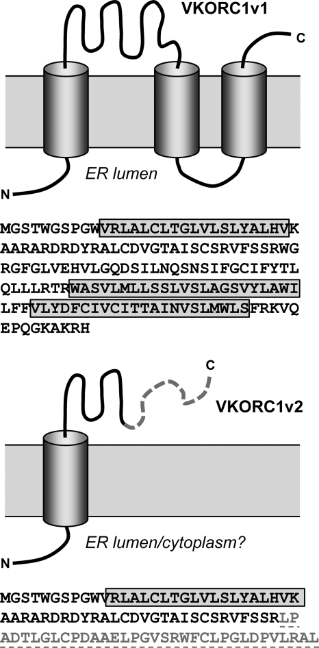
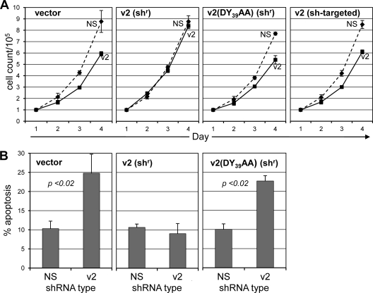
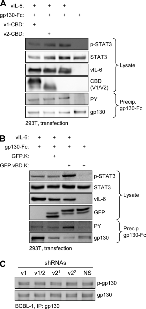
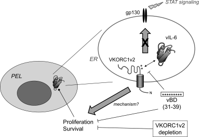
Viral interleukin-6 (vIL-6) specified by human herpesvirus 8 is, unlike its cellular counterpart, secreted very inefficiently and can signal via vIL-6(2):gp130(2) signaling complexes from the endoplasmic reticulum (ER) compartment. Intracellular, autocrine activities of vIL-6 are important for proproliferative and prosurvival activities of the viral cytokine in latently infected primary effusion lymphoma (PEL) cells. However, the molecular determinants of vIL-6 ER localization and function are unclear. Using yeast two-hybrid analysis, we identified the database-documented but uncharacterized splice variant of vitamin K epoxide reductase complex subunit 1 (VKORC1), termed VKORC1 variant 2 (VKORC1v2), as a potential interaction partner of vIL-6. In transfected cells, epitope-tagged VKORC1v2 was found to localize to the ER, to adopt a single-transmembrane (TM) topology placing the C tail in the ER lumen, and to bind vIL-6 via these sequences. Deletion mutagenesis and coprecipitation assays mapped the vIL-6-binding domain (vBD) of VKORC1v2 to TM-proximal residues 31 to 39. However, while sufficient to confer vIL-6 binding to a heterologous protein, vBD was unable to induce vIL-6 secretion when fused to (secreted) hIL-6, suggesting a VKORC1v2-independent mechanism of vIL-6 ER retention. In functional assays, overexpression of ER-directed vBD led to suppression of PEL cell proliferation and viability, effects also mediated by VKORC1v2 depletion and, as reported previously, by vIL-6 suppression. The growth-inhibitory and proapoptotic effects of VKORC1v2 depletion could be rescued by transduced wild-type VKORC1v2 but not by a vIL-6-refractory vBD-altered variant, indicating the functional relevance of the vIL-6-VKORC1v2 interaction. Notably, gp130 signaling was unaffected by VKORC1v2 or vBD overexpression or by VKORC1v2 depletion, suggesting an alternative pathway of vIL-6 activity via VKORC1v2. Combined, our data identify a novel and functionally significant interaction partner of vIL-6 that could potentially be targeted for therapeutic benefit.


Figures

Fig 1
Fig 1
Relationship between VKORC1 variants 1 and 2. Sequences and diagrammatic representations of the predicted structures of VKORC1 variants 1 and 2 are shown. The membrane topology shown for VKORC1v1 corresponds to that deduced by Tie et al. (21). Variant 2 shares the first 58 amino acids with variant 1; residues 59 to 92 are unique.
Fig 2
Fig 2
Interaction between vIL-6 and VKORC1v2. Association of vIL-6 with VKORC1v2 was identified by coprecipitation of vIL-6 with chitin bead-precipitated VKORC1v2-CBD from lysates of transfected HEK293T cells. Precipitated proteins (Ppt) were identified by immunoblotting (IB) for detection of vIL-6 and CBD. The two bands for vIL-6 represent glycosylated and unglycosylated forms of the protein. VKORC1v1-CBD did not precipitate vIL-6 (left). The right panels present the results of an analogous experiment, showing specificity of VKORC1v2 binding to vIL-6; no interaction between VKORC1v2 and ER protein calreticulin (Crt) was detected. vec, empty vector.
Fig 3
Fig 3
ER localization and membrane topology of VKORC1v2. (A) Confocal immunofluorescence assay results, showing colocalization of ER-directed GFP-KDEL and Flag-tagged VKORC1v2 in transfected HEK293T cells. (B) Confocal immunofluorescence-determined colocalization of vIL-6 and VKORC1v2-Flag in transfected HEK293T cells (two fields shown). (C) Immunoblot detection of VKORC1v2 in sucrose gradient-purified ER membrane preparations derived from VKORC1v2-Flag-transfected HEK293T cells, confirming ER localization of VKORC1v2. Calreticulin (Crt; ER localized) and early endosome antigen 1 (EEA1) provided quality control markers for ER preparations. (D) Determination of VKORC1v2 orientation within the ER by N-glycosylation tagging, showing efficient (∼100%) glycosylation of variant 2 upon introduction of an N-glysosylation site (N45GT by V45N mutation) into the post-TM region. These results demonstrate lumenal exposure of this region. In contrast, no such glycosylation was observed for VKORC1v1 containing the identical introduced N-glycosylation site. Controls for endo H digestion and resistance to digestion were provided by vIL-6 (high mannose glycosylated) and gp130 (containing mature glycan moieties), respectively. (E) Protease digestion experiment using C-terminally CBD-tagged VKORC1v2 revealed protection of CBD (and most/all of VKORC1v2) upon exposure of transfected cell-derived ER membrane preparations to proteinase K (Prot. K). The cytoplasmically exposed CBD of C-terminally tagged calnexin (ER transmembrane protein) and lumenally expressed calreticulin were cleaved and protected, respectively, verifying protease activity and membrane integrity.
Fig 4
Fig 4
Mapping of VKORC1v2 residues interacting with vIL-6. (A) Coprecipitation assays were employed to identify the region(s) of VKORC1v2 required for its interaction with vIL-6; CBD-tagged VKORC1v2 and deleted derivatives were utilized in chitin-mediated precipitations (IP) from transfected cell lysates. The region of VKORC1v2 immediately following the TM domain was found to be required for binding. Dialanine mutations within this region (residues 31 to 39) were introduced into VKORC1v2-CBD and tested for interaction with vIL-6; the DY39AA mutation inhibited binding. IB, immunoblotting; WT, wild type. (B) Residues 31 to 39, constituting the vIL-6 binding domain of VKORC1v2, were transferred to a heterologous protein, KDEL-tagged hIL-6, to test their sufficiency for binding to vIL-6. The nonopeptide sequence was able to confer vIL-6 binding to hIL-6-KDEL in transfected cells. (C) In vitro coprecipitation assay using media-derived vIL-6–CBD and bacterially expressed thioredoxin (Trx)/His6-fused VKORC1v2 C tail (residues 31 to 92), demonstrating direct interaction between the two proteins. Trx-His6 (negative control) was unable to bind vIL-6–CBD, and VKORC1v2 protein could not be coprecipitated with control hIL-6-CBD.
Fig 5
Fig 5
VKORC1v2 in intracellular retention of vIL-6. (A) The influence of VKORC1v2 on intracellular retention of vIL-6 was tested by overexpression of vBD as a fusion with hIL-6 (secreted) or hIL-6-KDEL (ER retained) in vIL-6-cotransfected HEK293T cells. While ER-expressed vBD led to a reduction in the very low levels of secreted vIL-6, consistent with its binding the viral cytokine, vBD in the context of secreted hIL-6 was insufficient to induce, in a dominant fashion, secretion of vIL-6. These data indicate that a mechanism independent of vIL-6–VKORC1v2 association is responsible for ER retention of vIL-6. (B) The ability of hIL-6-fused vBD to disrupt vIL-6–VKORC1v2 binding was verified in a coprecipitation assay utilizing lysates from transfected HEK293T cells. Chitin bead-sedimented VKORC1v2-CBD was able to coprecipitate vIL-6 in the presence of hIL-6-KDEL (control) but not its vBD-containing counterpart. (C) Testing by RT-PCR (top panels) of lentivirus-cloned shRNAs directed to VKORC1 variants 1 and/or 2 transcripts (see Materials and Methods) for their abilities to deplete the respective mRNAs in BCBL-1 cells. Selected, active shRNAs were then tested in VKORC1v1- and VKORC1v2-vector transfected cells to verify by immunoblotting (IB; bottom panels) their activities in respect of VKORC1 protein depletion. (D) shRNA-mediated VKORC1v2 depletion had little effect relative to controls (VKORC1v1 and NS shRNA transduction) on the levels of secreted, medium-derived versus intracellular vIL-6. Secreted vIL-6 was immunoprecipitated from medium, and the proportion loaded onto the gel and immunoblotted represents 50 times the fraction of corresponding cell lysate analyzed. (E) Western analysis of density gradient-purified ER membrane preparations by immunoblotting revealed that there was no influence of VKORC1v2 depletion on ER localization of vIL-6. Calreticulin (Crt; ER marker) and EEA1 (endosomal marker) were detected to check the quality of the ER preparations.
Fig 6
Fig 6
Functional analysis of VKORC1v2 and the vIL-6–VKORC1v2 interaction. (A) Lentivirus-shRNA transduction of PEL cells revealed substantial inhibition of cell growth by VKORC1v2-directed shRNAs (v2 alone or v1+2) relative to NS control and v1-targeted shRNAs. Growth of HHV-8-negative BJAB cells was unaffected or minimally altered by transduction of these VKORC1 shRNAs. JSC-1 PEL cell growth, like that of BCBL-1 cells, was inhibited markedly in response to VKORC1v2-specific shRNA, while control Akata cells (HHV-8 negative) were unaffected. (B) Lentivirus vector-mediated expression in BCBL-1 and JSC-1 cells of ER-directed vBD (GFP-vBD-KDEL), used as a competitor for the VKORC1v2–vIL-6 interaction, also led to growth inhibition. This was not seen in identically treated BJAB and Akata cells. (C) Individual and cotransduction of v2-shRNA and vBD in BCBL-1 cells failed to reveal significant additive effects. These data are consistent with targeting of a common mechanism rather than distinct pathways. (D) Using Annexin V-Cy3 staining for identification of apoptotic cells, both VKORC1v2 depletion and GFP-vBD-KDEL (vBD-K) transduction were found to lead to significantly increased rates of BCBL-1 cell apoptosis relative to those detected in NS-shRNA and GFP-KDEL (K) control cultures (left and right panels, respectively). For all experiments (panels A to D), triplicate cultures were used; error bars represent standard deviations from mean values. In panel D, NS versus v2 P values (t test, unpaired, two tailed) are shown.
Fig 7
Fig 7
Role of the VKORC1–vIL-6 interaction in PEL cell growth and survival. (A) BCBL-1 cells were coinfected with lentivirus vectors specifying control (NS) or VKORC1v2-specific shRNA and “shRNA-resistant” (shr) wild-type or DY39AA-mutated (vIL-6-refractory) VKORC1v2 proteins, or wild-type VKORC1v2 susceptible to shRNA targeting (control for effective depletion). Cell growth was monitored daily following lentivirus transduction and a 2-day rest by counting of trypan blue-excluding cells. (B) In a parallel experiment, apoptosis was assayed at day 4 based on Annexin V-Cy3 staining and calculated as the fraction of total cells in each of >10 random fields. For growth and apoptosis data, error bars represent deviations from mean values obtained from triplicate cultures; t test values (unpaired, two tailed) are shown in panel B.
Fig 8
Fig 8
Testing the potential influence of VKORC1v2 on vIL-6 signaling via gp130. (A) Cotransfection assays were undertaken to determine the effect of VKORC1v2 (and VKORC1v1 [control]) overexpression on vIL-6/gp130 signaling, induced by coexpression of vIL-6 and (functional) gp130-Fc. Immunoblotting was used to identify phospho-STAT3 (p-STAT3) in cell lysates and tyrosine-phosphorylated (PY) gp130-Fc following precipitation from cell lysates by protein A-agarose as measures of vIL-6-induced gp130 signaling and activation. VKORC1v2 had no effect on either. (B) Overexpression of vBD, fused to GFP-KDEL, with vIL-6-KDEL (fully ER retained) and gp130-Fc, had no inhibitory effect on STAT3 activation or gp130 phosphorylation but rather seemed to enhance vIL-6 activity. (C) Depletion of VKORC1v2 in BCBL-1 cells did not alter the levels of active (phosphorylated) endogenous gp130, immunoprecipitated from cell lysate prior to immunoblotting for detection of phospho-tyrosine (p-gp130).
Fig 9
Fig 9
Summary of our main findings and conclusions. Previous studies revealed that vIL-6 is expressed in latently infected PEL cells and functions in the ER compartment to promote cell growth and survival (5). The present study identified VKORC1v2 as an ER-localized binding partner of vIL-6 and mapped the vIL-6-binding region of VKORC1v2 to residues 31 to 39 (vBD), which is capable, when fused to a heterologous protein and targeted to the ER, of interfering with vIL-6 binding to VKORC1v2 and suppressing PEL growth and survival. These outcomes were also seen upon VKORC1v2 depletion and could be rescued by (shRNA-refractory) wild-type VKORC1v2 but not by a dialanine variant (DY39AA) with diminished ability to bind vIL-6. Neither VKORC1v2 depletion nor vBD overexpression had detectable negative effects on gp130 signaling, and VKORC1v2 overexpression did not enhance it. These findings suggest that activities of vIL-6 connected with its binding to VKORC1v2 are mediated independently of g130 signaling, which is known to promote growth and survival of various cell types. However, the mechanism of vIL-6 activity via VKORC1v2 association remains to be determined.

References

    1. Adam N, et al. 2009. Unraveling viral interleukin-6 binding to gp130 and activation of STAT-signaling pathways independently of the interleukin-6 receptor. J. Virol. 83:5117–5126 - PMC - PubMed
    1. Cain D, Hutson SM, Wallin R. 1997. Assembly of the warfarin-sensitive vitamin K 2,3-epoxide reductase enzyme complex in the endoplasmic reticulum membrane. J. Biol. Chem. 272:29068–29075 - PubMed
    1. Chen D, Choi YB, Sandford G, Nicholas J. 2009. Determinants of secretion and intracellular localization of human herpesvirus 8 interleukin-6. J. Virol. 83:6874–6882 - PMC - PubMed
    1. Chen D, Nicholas J. 2006. Structural requirements for gp80 independence of human herpesvirus 8 interleukin-6 (vIL-6) and evidence for gp80 stabilization of gp130 signaling complexes induced by vIL-6. J. Virol. 80:9811–9821 - PMC - PubMed
    1. Chen D, Sandford G, Nicholas J. 2009. Intracellular signaling mechanisms and activities of human herpesvirus 8 interleukin-6. J. Virol. 83:722–733 - PMC - PubMed

Publication types

LinkOut - more resources