. 2012 Feb;86(4):2259-72.
doi: 10.1128/JVI.05906-11. Epub 2011 Nov 30.

The tetherin/BST-2 coiled-coil ectodomain mediates plasma membrane microdomain localization and restriction of particle release

Affiliations

The tetherin/BST-2 coiled-coil ectodomain mediates plasma membrane microdomain localization and restriction of particle release

Jason Hammonds et al. J Virol. 2012 Feb.

Abstract

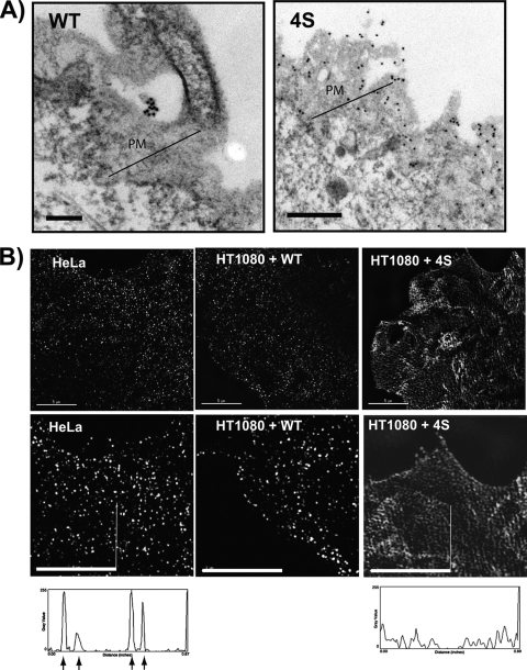
Tetherin/BST-2 forms a proteinaceous tether that restricts the release of a number of enveloped viruses following viral budding. Tetherin is an unusual membrane glycoprotein with two membrane anchors and an extended coiled-coil ectodomain. The ectodomain itself forms an imperfect coil that may undergo conformational shifts to accommodate membrane dynamics during the budding process. The coiled-coil ectodomain is required for restriction, but precisely how it contributes to the restriction of particle release remains under investigation. In this study, mutagenesis of the ectodomain was used to further define the role of the coiled-coil ectodomain in restriction. Scanning mutagenesis throughout much of the ectodomain failed to disrupt the ability of tetherin to restrict HIV particle release, indicating a high degree of plasticity. Targeted N- and C-terminal substitutions disrupting the coiled coil led to both a loss of restriction and an alteration of subcellular distribution. Two ectodomain mutants deficient in restriction were endocytosed inefficiently, and the levels of these mutants on the cell surface were significantly enhanced. An ectodomain mutant with four targeted serine substitutions (4S) failed to cluster in membrane microdomains, was deficient in restriction of particle release, and exhibited an increase in lateral mobility on the membrane. These results suggest that the tetherin ectodomain contributes to microdomain localization and to constrained lateral mobility. We propose that focal clustering of tetherin via ectodomain interactions plays a role in restriction of particle release.


Figures

Fig 1
Fig 1
Alanine scanning mutagenesis of the human tetherin ectodomain. HEK293T cells were transfected with 100 ng of FLAG-tagged tetherin expression plasmids and 1 μg of pNL4.3/Udel (NLUdel). At 24 h posttransfection, cells and supernatants were collected and processed for Western blot analysis. (A) Schematic representation of tetherin ectodomain alanine scanning mutagenesis. FLAG, N-terminal FLAG tag; CD, cytoplasmic domain, TM, transmembrane domain; GPI, glycosyl-phosphatidylinositol anchor. (B) Viral and cell lysate protein detection using anti-p24 and tetherin specific antibodies. Numbers above the blots correspond to the position of alanine substitutions. Sup, particles pelleted from supernatants.
Fig 2
Fig 2
Targeted serine substitutions within the tetherin coiled-coil region exhibit loss of HIV particle retention. HEK293T cells were transfected with 100 ng of FLAG-tagged tetherin expression plasmids and 1 μg of NLUdel. At 24 h posttransfection, cells and supernatants were collected and processed for Western blot analysis. (A) Schematic representation of tetherin ectodomain serine substitution mutations. (B) Viral and cell lysate protein detection using anti-p24 and tetherin specific antibodies. NLUdel alone (no tetherin, leftmost lane) and NLUdel + wild-type tetherin (second lane from left) served as controls for no restriction and wild-type restriction, respectively. (C) Infectivity measurement of pelleted particles from tetherin and NLUdel-transfected cells. Transfection of increasing amount of tetherin is shown on the x axis, with relative light units measured in the TZM-bl reporter cell line on the y axis. (D) Analysis of dimer formation by tetherin serine mutants under nonreducing (left four lanes) and reducing conditions (right four lanes). Position of tetherin monomers versus dimers is indicated on the right.
Fig 3
Fig 3
Tetherin ectodomain mutants display altered cell surface expression. (A) Structure of coiled-coil ectodomain (from Schubert et al. [33]) for reference to alanine substitution positions. Positions altered in serine mutants designed to disrupt hydrophobic interactions are shown in red and bolded. (B) Cell surface proportion of tetherin constructs. 293T cells were cotransfected with the indicated flag-tagged tetherin expression construct together with a GFP-expression plasmid. At 24 h posttransfection, the cells were harvested, and GFP+ were cells analyzed for tetherin cell surface expression by flow cytometry. Both cell surface (nonpermeabilized) and total tetherin expression was measured; cell surface expression was normalized to total tetherin expression in permeabilized cells, with the wild type set at 1.0. Error bars represent the standard deviation of three separate experiments. (C) FACS plots of wild-type and serine mutant cell surface expression. The dotted plot represents the isotype control, the gray plot is the wild-type cell surface expression, and the dark unfilled plot represents the mutants indicated.
Fig 4
Fig 4
Endocytosis rates of tetherin and tetherin ectodomain mutants. HEK293T cells were cotransfected with the indicated tetherin expression construct and a GFP expression plasmid. At 24 h posttransfection, the cells were detached with EDTA and surface labeled with rabbit anti-tetherin antisera at 4°C. Cell aliquots were incubated at 37°C for the indicated time intervals in order to facilitate endocytosis. Cells were then rapidly cooled to 4°C and stained with the appropriate secondary antibody. Relative surface tetherin levels in GFP+ cell populations were set to 100% at time zero. Error bars represent results from three concurrent experiments. Wild-type tetherin is indicated by filled squares and the dashed line.
Fig 5
Fig 5
Subcellular distribution of tetherin ectodomain mutants. HT1080 cells were transfected with wild-type (A), 79.82A (B), 4S (C), and 135-138A (D) tetherin expression plasmids. At 20 h posttransfection, cells were fixed, permeabilized, and costained with anti-tetherin (green), anti-TGN46 (red), and nuclei with DAPI (blue). Cells were visualized using wide-field fluorescence deconvolution microscopy on a Deltavision imaging station (Applied Precision). The cells shown for each construct are representative of 100 cells examined.
Fig 6
Fig 6
Immunoelectron and super-resolution microscopic analysis of plasma membrane tetherin distribution. (A) HT1080 cells grown on coverslips were fixed and stained with anti-tetherin antibody and anti-rabbit immunogold beads prior to further fixation and sectioning. Sectioning was performed tangential to the plasma membrane as described in Materials and Methods. Wild-type (WT) tetherin cluster on cellular extension, representative of focal clustering. 4S distribution on plasma membrane demonstrates the higher expression and diffuse nature of 4S tetherin. Scale bars, 200 nm. (B) Super-resolution microscopic images of tetherin on the plasma membrane of HT1080 cells were gathered using structured illumination microscopy. Cells were stained with anti-tetherin antisera in the absence of permeabilization, and a z-stack of images gathered on the OMX structured illumination imaging station version 3 (Applied Precision). Image acquisition and reconstruction was performed with the SoftWorx software package from Applied Precision, and an individual section at the cell surface is shown in the panels at two different magnifications. Endogenous tetherin in HeLa cells is shown in leftmost panels. HT1080 cells expressing WT tetherin are shown in middle panels, and HT1080 cells with 4S tetherin are shown on the right. Vertical bars on leftmost and rightmost panels indicate lines used to generate line plots of signal intensity shown below corresponding images. Arrows indicate peaks representing WT tetherin clusters. Scale bars, 5 μm.
Fig 7
Fig 7
WT and 4S tetherin colocalization with clathrin-GFP, Gag, and subcellular localization of additional tetherin mutants. (A) Clathrin-GFP (green) and plasma membrane tetherin (red) were examined at the periphery of transfected HT1080 cells for punctate colocalization. Square in leftmost image indicates area examined for colocalization. Colocalized puncta are shown in rightmost panel. After appropriate thresholding for each wavelength to measure only those pixels associated with intense puncta, a colocalization coefficient of 0.84 was obtained (green puncta colocalizing with red puncta). Size bars: 11 μm (far left images) and 2.7 μm (right three images). (B) Clathrin-GFP and 4S tetherin colocalization. Diffuse nature of 4S precludes meaningful colocalization quantitation, but there was no obvious concentration of red pixels where green signal is concentrated. (C) HT1080 cells expressing NLUdel and WT tetherin were stained with anti-tetherin antisera and then permeabilized and stained with anti-Gag monoclonal antibody. Colocalized puncta on plasma membrane are indicated by arrows. Size bars, 5.4 μm. (D) HT1080 cells expressing 4S tetherin were treated as in panel C above. Location of Gag puncta reveals no concentration of 4S tetherin (red). Size bars, 5.4 μm. (E) Surface staining for tetherin comparing 4S, tetherin 71-74A, and tetherin 95-98A. Size bars, 11 μm.
Fig 8
Fig 8
Characterization of N-terminal GFP-tagged tetherin ectodomain mutants. HEK 293T cells were transfected with 100 ng of GFP-tagged tetherin expression plasmids and 1 μg of NLUdel. At 24 h posttransfection, the cells and supernatants were collected and processed for Western blot analysis. (A) Viral and cell lysate protein detection were performed using anti-p24 and GFP-specific antibodies. Pelleted NLUdel particles are shown below as p24 band. (B) Subcellular distribution of GFP-tetherin (WT) and GFP-4S tetherin was examined on a wide-field deconvolution imaging system. Size bars: 11 μm (left) and 16 μm (right).
Fig 9
Fig 9
Fluorescence recovery after photobleaching for wild-type and ectodomain mutant tetherin. HT1080 cells were transfected with 100 ng of indicated GFP-tagged tetherin expression plasmid. At 24 posttransfection a membrane region of interest (ROI) close to the coverslip was bleached using high intensity laser settings. Fluorescence recovery was measured every second for a total of 30 s postbleaching. (A) Representative image series for wild-type tetherin (top panels) and 4S tetherin (lower panels). A larger field view is at left. Dashed circle indicates targeted region for bleaching. The 1.0-s time point represents the first postbleaching image. Bleached area is indicated by dashed circle. Scale bar, 10 μm. (B) Representative FRAP curves are shown for wild-type and 4S tetherin. An arrow indicates the time of laser pulse. (C) From the fluorescence recovery half times, the diffusion coefficient was calculated from FRAP measurements of 10 cells each. Statistical significance was performed using an unpaired t test, comparing to WT levels. *, P < 0.05.

Similar articles

Cited by

References

    1. Andrew AJ, Miyagi E, Strebel K. 2011. Differential effects of human immunodeficiency virus type 1 Vpu on the stability of BST-2/tetherin. J. Virol. 85:2611–2619 - PMC - PubMed
    1. Axelrod D, Koppel DE, Schlessinger J, Elson E, Webb WW. 1976. Mobility measurement by analysis of fluorescence photobleaching recovery kinetics. Biophys. J. 16:1055–1069 - PMC - PubMed
    1. Chen WH, Da WM, Gao CJ. 2009. Construction of ICAM-1-GFP and its binding with Molt-4 cells. Zhongguo Shi Yan Xue Ye Xue Za Zhi. 17:650–655 - PubMed
    1. Cohen EA, Terwilliger EF, Sodroski JG, Haseltine WA. 1988. Identification of a protein encoded by the vpu gene of HIV-1. Nature 334:532–534 - PubMed
    1. Douglas JL, et al. 2010. The great escape: viral strategies to counter BST-2/tetherin. PLoS Pathog. 6:e1000913. - PMC - PubMed

Publication types