. 2012 Feb;86(3):1777-88.
doi: 10.1128/JVI.06626-11. Epub 2011 Nov 30.

Accelerated shedding of prions following damage to the olfactory epithelium

Affiliations

Accelerated shedding of prions following damage to the olfactory epithelium

Richard A Bessen et al. J Virol. 2012 Feb.

Abstract

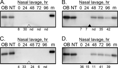
In this study, we investigated the role of damage to the nasal mucosa in the shedding of prions into nasal samples as a pathway for prion transmission. Here, we demonstrate that prions can replicate to high levels in the olfactory sensory epithelium (OSE) in hamsters and that induction of apoptosis in olfactory receptor neurons (ORNs) in the OSE resulted in sloughing off of the OSE from nasal turbinates into the lumen of the nasal airway. In the absence of nasotoxic treatment, olfactory marker protein (OMP), which is specific for ORNs, was not detected in nasal lavage samples. However, after nasotoxic treatment that leads to apoptosis of ORNs, both OMP and prion proteins were present in nasal lavage samples. The cellular debris that was released from the OSE into the lumen of the nasal airway was positive for both OMP and the disease-specific isoform of the prion protein, PrP(Sc). By using the real-time quaking-induced conversion assay to quantify prions, a 100- to 1,000-fold increase in prion seeding activity was observed in nasal lavage samples following nasotoxic treatment. Since neurons replicate prions to higher levels than other cell types and ORNs are the most environmentally exposed neurons, we propose that an increase in ORN apoptosis or damage to the nasal mucosa in a host with a preexisting prion infection of the OSE could lead to a substantial increase in the release of prion infectivity into nasal samples. This mechanism of prion shedding from the olfactory mucosa could contribute to prion transmission.


Figures

Fig 1
Fig 1
Disruption of the olfactory sensory epithelium following nasotoxic injury. The nasal turbinates in the nasal cavity from mock-infected hamsters treated with vehicle only (A and B) or methimazole (C, D, E, and F) were analyzed for OMP by using hematoxylin and eosin (H&E) (A and C) and immunohistochemistry (brown) (B and D) at 24 h after treatment. (B) OMP is prominent in the OSE and nerve bundles (NB) in the subepithelial layer (SE) in the absence of methimazole treatment. (C and D) Following methimazole treatment, the OSE was observed to slough off the nasal turbinates (arrowhead in panel C), but OMP immunostaining was still observed in nerve bundles of the SE (D). Some OMP immunoreactivity was observed in cellular debris in the lumen of the nasal airway (arrowhead in panel D). In panels C and D, the labels E and F refer to areas that were enlarged and illustrated in panels E and F. NA, lumen of the nasal airway.
Fig 2
Fig 2
Apoptotic index for nasal turbinates following methimazole treatment. The apoptotic index was measured as the ratio of cl-caspase 3 to total caspase 3 (A) and the ratio of cl-caspase 3 to actin (B) by Western blotting as described in Materials and Methods. Postmethimazole (post-MTz) is the time point in hours at which nasal turbinates were collected for cl-caspase 3, pro-caspase 3, and actin analyses after methimazole treatment. Asterisks indicate a P value of <0.001 (unpaired t test) for comparison of the vehicle group to each methimazole group.
Fig 3
Fig 3
Distribution of cleaved caspase 3 following apoptosis of olfactory receptor neurons. Hamsters were treated with either vehicle alone (A) or methimazole (B, C, and D), and the nasal turbinates (A and B) and olfactory bulb (C and D) were collected 12 and 24 h following treatment for analysis of apoptosis by cleaved caspase 3 immunofluorescence (red). Cleaved caspase 3 was infrequent in the OSE and olfactory bulb samples from vehicle-treated hamsters (A and data not shown). Following methimazole treatment, cl-caspase 3 immunofluorescence was observed in the disrupted OSE (arrows in panel B) in a pattern consistent with deposition in ORNs, as well as in the outer nerve layer (ONL) and glomeruli (*) in the olfactory bulb (C and D). The ONL is composed of the axons of the ORNs, and the glomeruli are synapse-rich structures containing the nerve terminals of ORNs as well as dendrites from neurons in the olfactory bulb. An increase in cl-caspase 3 in the OSE, ONL, and glomeruli is consistent with apoptosis of ORNs following methimazole treatment. Nuclei are stained with ToPro 3 (blue). SE, subepithelial layer; NA, lumen of the nasal airway. Scale bars, 50 μm.
Fig 4
Fig 4
Western blots for OMP in olfactory tissues and nasal lavage samples following nasotoxic injury. Age-matched mock-infected (A and B) and HY TME agent-infected (C and D) hamsters were treated with vehicle only (A and C) or methimazole (B and D) after the collection of nasal lavage samples at the 24-h time point (open and filled arrowheads, respectively). Nasal lavage samples were collected every 24 h for five consecutive days, and after the 96-h collection point, the olfactory bulb (OB) and nasal turbinate (NT) were also collected for OMP analysis. Fifty micrograms of protein for the OB and NT lysates and 200 μl of nasal lavage fluid were analyzed by Western blotting using polyclonal anti-OMP goat antibody. The total amount of protein in nasal lavage samples varied from below the level of detection (i.e., <1 μg/ml) to 70 μg protein. The value under each nasal lavage lane indicates the total amount of protein (in micrograms) analyzed. Nd refers to none detected and indicates levels below the limit of detection. The marker (m) lane contains a polypeptide at 20 kDa.
Fig 5
Fig 5
Distribution of OMP and PrPSc following disruption of the olfactory sensory epithelium. The olfactory sensory epithelium in the nasal cavity from mock-infected (A to F) and HY TME agent-infected (G to L) hamsters was analyzed by laser scanning confocal microscopy for OMP (red) (A, D, G, and J), PrPSc (green) (B, E, H, and K), and for both OMP and PrPSc (C, F, I, and L). Panels A through C, D through F, G through I, and J through L show the same fields of view. Hamsters were treated either with vehicle alone (A to C and G to I) or with methimazole (D to F and J to L) and analyzed 72 h after treatment. OMP is present in olfactory receptor neurons, and immunofluorescence was observed in the OSE (the white bar indicates the width of the OSE) and nerve bundles (*) in the vehicle-treated group and primarily in the nerve bundles following methimazole treatment. OMP immunofluorescence in the OSE was less frequent following methimazole treatment and disruption of the OSE, but it could be observed separating from the epithelium (arrow in panels D, F, J, and L) and in the lumen of the nasal airway (NA). PrPSc was not observed in mock-infected hamsters but was present in the OSE of HY TME hamsters in the vehicle group (H) and in the disrupted OSE of the methimazole-treated HY TME hamsters (arrow in panels K and L). The nuclei (blue) of olfactory receptor neurons are packed at a high density in the OSE of the vehicle-treated groups. Scale bar, 50 μm.
Fig 6
Fig 6
Western blots for prion protein in olfactory tissues and nasal lavage samples following nasotoxic injury. Age-matched mock-infected (A and B) and HY TME agent-infected (C and D) hamsters were treated with vehicle only (A and C) or methimazole (B and D) after the collection of nasal lavage samples at the 24-h time point (open and filled arrowheads, respectively). Nasal lavage samples were collected every 24 h for five consecutive days, and after the 96-h collection point, the olfactory bulb (OB) and nasal turbinate (NT) were also collected for PrP analysis. For the OB and NT lysates, 10 μg (B and D), 12.5 μg (C), or 50 μg (A) of protein was analyzed while for the nasal lavage samples 200 μl was analyzed by Western blotting using monoclonal anti-PrP 3F4 mouse antibody. The total amount of protein in nasal lavage samples varied from below the level of detection (i.e., <1 μg/ml) to 70 μg protein. The marker (m) lane contains polypeptides at 20, 30, and 40 kDa.
Fig 7
Fig 7
RT-QuIC assay of nasal lavage samples from HY TME hamsters. The time to maximum ThT fluorescence in the RT-QuIC assay was measured for nasal lavage samples collected every 24 h for five consecutive days from HY TME hamsters following treatment with vehicle only (open bars) or methimazole (closed bars), which was administered after the collection of nasal lavage samples at the 24-h time point. Nasal lavage samples from three to five hamsters were assayed at each time point for both treatment groups. Samples that did not reach maximum ThT fluorescence by 63 h were not included in the analysis, but this set consisted only of samples from either the vehicle or pre-methimazole treatment group. At each collection point, the values for the vehicle and methimazole groups were compared using a paired t test (two-tailed). *, P < 0.01; **, P < 0.001.
Fig 8
Fig 8
RT-QuIC assay of olfactory tissues from HY TME agent-infected hamsters. The RT-QuIC assay was used to measure ThT fluorescence for a duration of 63 h in individual reactions seeded with serial dilutions of olfactory bulb samples (A), nasal turbinate samples (B), nasal lavage samples from the 0-h collection point (T0) (C), and nasal lavage samples from the 48-h collection point (T48; 24 h after methimazole treatment) (D). Samples were assayed neat and serially diluted 10-fold (10-f) to 1,000-fold (1K-f), 1,000,000-fold (1 M-f), and 1,000,000,000-fold (1B-f). Normal hamster brain (NBH) was used as a negative control and 100 fg of PrPSc from a 263K scrapie strain-infected hamster brain was used as a positive control in the RT-QuIC assay. Four replicate wells were assayed for each sample, and each data point on the curve is the average of results for replicates. The corresponding median prion seeding dose (SD50) for these tissues (H1636.3) can be found in Table 2. Panels representative of results from the RT-QuIC assay are shown.

Similar articles

Cited by

References

    1. Apter AJ, Gent JF, Frank ME. 1999. Fluctuating olfactory sensitivity and distorted odor perception in allergic rhinitis. Arch. Otolaryngol. Head Neck Surg. 125:1005–1010 - PubMed
    1. Atarashi R, et al. 2008. Simplified ultrasensitive prion detection by recombinant PrP conversion with shaking. Nat. Methods 5:211–212 - PubMed
    1. Bahrami F, van Hezik C, Bergman A, Brandt I. 2000. Target cells for methylsulphonyl-2,6-dichlorobenzene in the olfactory mucosa in mice. Chem. Biol. Interact. 128:97–113 - PubMed
    1. Balkema-Buschmann A, et al. 2011. BSE infectivity in the absence of detectable PrP(Sc) accumulation in the tongue and nasal mucosa of terminally diseased cattle. J. Gen. Virol. 92:467–476 - PubMed
    1. Bartz JC, Kincaid AE, Bessen RA. 2003. Rapid prion neuroinvasion following tongue infection. J. Virol. 77:583–591 - PMC - PubMed

Publication types