. 2012 Feb;86(4):1930-41.
doi: 10.1128/JVI.06127-11. Epub 2011 Nov 30.

Murine gammaherpesvirus 68 evades host cytokine production via replication transactivator-induced RelA degradation

Affiliations

Murine gammaherpesvirus 68 evades host cytokine production via replication transactivator-induced RelA degradation

Xiaonan Dong et al. J Virol. 2012 Feb.

Abstract

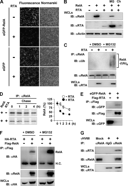
Cytokines play crucial roles in curtailing the propagation and spread of pathogens within the host. As obligate pathogens, gammaherpesviruses have evolved a plethora of mechanisms to evade host immune responses. We have previously shown that murine gammaherpesvirus 68 (γHV68) induces the degradation of RelA, an essential subunit of the transcriptionally active NF-κB dimer, to evade cytokine production. Here, we report that the immediately early gene product of γHV68, replication transactivator (RTA), functions as a ubiquitin E3 ligase to promote RelA degradation and abrogate cytokine production. A targeted genomic screen identified that RTA, out of 24 candidates, induces RelA degradation and abolishes NF-κB activation. Biochemical analyses indicated that RTA interacts with RelA and promotes RelA ubiquitination, thereby facilitating RelA degradation. Mutations within a conserved cysteine/histidine-rich, putative E3 ligase domain impaired the ability of RTA to induce RelA ubiquitination and degradation. Moreover, infection by recombinant γHV68 carrying mutations that diminish the E3 ligase activity of RTA resulted in more robust NF-κB activation and cytokine induction than did infection by wild-type γHV68. These findings support the conclusion that γHV68 subverts early NF-κB activation and cytokine production through RTA-induced RelA degradation, uncovering a key function of RTA that antagonizes the intrinsic cytokine production during gammaherpesvirus infection.


Figures

Fig 1
Fig 1
Identification of γHV68 RTA that abrogates NF-κB activation. (A) 293T cells were transfected with a reporter plasmid cocktail and a plasmid containing the individual γHV68 gene. NF-κB activation by RelA was assessed by a luciferase assay. I.E., immediate-early. The P value was calculated in reference to transfection with a plasmid containing RelA alone. (B) Key components and their relation in NF-κB signaling pathways that lead to cytokine production. (C) 293T cells were transfected with a reporter plasmid cocktail and with plasmids containing the indicated host genes and γHV68 RTA. The relative fold change by the coexpression of RTA is shown in reference to the vector (Vec). The P value is <0.02 for all pairs. (D) T-REx 293 cell lines carrying the vector or γHV68 RTA were preincubated with doxycycline (DOX) (1 μg/ml) for 24 h and transfected with a reporter plasmid cocktail and a plasmid containing TRAF6. NF-κB activation was assessed by a luciferase reporter assay. (E) γHV68 RTA mRNA levels in T-REx 293 cell lines carrying the vector or γHV68 RTA without or with doxycycline were assessed by real-time PCR. The relative quantity of RTA transcripts was normalized to that of β-actin. (F) 293 T-REx.Vec and 293 T-REx.mRTA cells were induced by doxycycline (1 μg/ml) for 24 h and infected with Sendai virus (SeV) for 8 h. Total RNA was extracted and analyzed by real-time PCR with primers specific for TNF-α and CXCL1. Data in panels A, C, D, and E are presented as the means ± standard errors of the means (SEM) from at least three independent experiments. The statistical significance (P value) was calculated by unpaired two-tailed Student's t test. **, P < 0.02.
Fig 2
Fig 2
γHV68 RTA promotes ubiquitination and degradation of RelA. (A) 293T cells were transfected with plasmids containing RTA and eGFP-tagged RelA or eGFP. eGFP- and eGFP-RelA-expressing cells were visualized by fluorescence microscopy (left) and Nomarski microscopy (right). (B) 293T cells were transfected with plasmids containing RelA and RTA. At 30 h posttransfection, cells were treated with MG132 (MG) (20 μM) or chloroquine (Ch) (50 μM) for 6 h. Whole-cell lysates (WCLs) were analyzed by immunoblotting (IB). (C) 293T cells were transfected with a vector or a plasmid containing γHV68 RTA. At 30 h posttransfection, cells were treated with MG132 (20 μM) for 6 h. WCLs were precipitated with anti-RelA antibody and analyzed by immunoblotting with anti-ubiquitin and anti-RelA antibodies. WCLs were analyzed by immunoblotting with anti-RTA antibody. (D) 293T cells were transfected with plasmids containing RelA and γHV68 RTA. At 36 h posttransfection, cells were pulse-labeled with [35S]methionine-cysteine for 30 min, chased, and harvested at the indicated time points. Precipitated RelA was analyzed by autoradiography. (E) 293T cells were transfected with plasmids containing eGFP-RelA and Flag-RTA. Centrifuged cell lysates were precipitated with anti-Flag antibody and analyzed by immunoblotting with antibodies to GFP and the Flag epitope. WCLs were analyzed by immunoblotting with anti-GFP antibody. (F) 293T cells were transfected with plasmids containing Flag-RelA and HA-RTA. At 48 h after transfection, cells were treated with dimethyl sulfoxide (DMSO) or MG132 (20 μM) for 2 h. Whole-cell lysates were precipitated with anti-Flag (RelA) antibody and analyzed by immunoblotting with anti-HA (RTA) and anti-RelA antibodies. Whole-cell lysates were analyzed by immunoblotting for RTA expression with an anti-HA antibody. Note that the correspondingly paired samples under DMSO- and MG132-treated conditions were processed on the same membranes. (G) MEFs were infected with γHV68 K3/GFP at a multiplicity of infection (MOI) of 20 for 16 h. Centrifuged cell lysates were precipitated with anti-RelA antibody or control rabbit IgG (rIgG) and analyzed by immunoblotting with antibodies to RTA and RelA. WCLs were analyzed by immunoblotting with anti-RTA antibody.
Fig 3
Fig 3
A conserved ubiquitin E3 ligase activity is necessary for γHV68 RTA to induce RelA ubiquitination and degradation. (A) The ubiquitin E3 ligase domain is highly conserved between γHV68 RTA and KSHV RTA. Two solid triangles mark the cysteine residues that are necessary for the ubiquitin E3 ligase activity of KSHV RTA. (B) 293T cells were transfected with vector (−) or a plasmid containing γHV68 wild-type RTA (WT) or the RTA C/S variant (C/S). At 30 h posttransfection, cells were treated with MG132 (20 μM) for 6 h. Whole-cell lysates (WCLs) were precipitated with anti-RelA antibody and analyzed by immunoblotting with anti-ubiquitin and anti-RelA antibodies. WCLs were analyzed by immunoblotting with anti-RTA antibody. (C) 293T cells were transfected with plasmids carrying the indicated genes. At 48 h after transfection, cells were treated with DMSO or MG132 (20 μM) for 2 h, and whole-cell lysates were prepared for immunoprecipitation with anti-Flag (RTA). The immunoprecipitated complexes were analyzed by immunoblotting with anti-HA (ubiquitin) and anti-RTA antibodies. (D) 293T cells were transfected with plasmids containing RelA and γHV68 wild-type RTA or the RTA C/S variant. At 36 h posttransfection, cells were pulse-labeled with [35S]methionine-cysteine for 30 min, chased, and harvested at the indicated time points. Precipitated RelA was analyzed by autoradiography, and data represent data from three independent experiments. (E) 293T cells were transfected with 200 ng plasmid containing GST and different doses (100 ng, 200 ng, and 500 ng) of a plasmid containing γHV68 wild-type RTA or the RTA C/S variant. WCLs were analyzed by immunoblotting with antibodies to GST and RTA. The relative RTA protein levels were calculated by normalization to GST protein levels. (F) 293T cells were transfected with plasmids containing RelA, Flag-tagged wild-type RTA, or the RTA C/S variant. Centrifuged cell lysates were precipitated with anti-Flag antibody and analyzed by immunoblotting with antibodies to RelA and RTA. WCLs were analyzed by immunoblotting with anti-RelA antibody.
Fig 4
Fig 4
Reduced ability of the RTA C/S variant to inhibit NF-κB activation. (A) 293T cells were transfected with plasmids containing RelA and wild-type RTA (WT) or the RTA C/S variant (C/S). At 20 h posttransfection, cells were treated with MG132 (20 μM) for 6 h or left untreated. WCLs were analyzed by immunoblotting. C/Sx3, 3-fold the amount of a plasmid carrying the RTA C/S variant was used, compared with other groups. The relative RTA and RelA protein levels were calculated by normalization to actin protein levels. (B) 293T cells were transfected with a reporter plasmid cocktail and plasmids containing RelA, wild-type RTA, or the RTA C/S variant. NF-κB activation was quantitated by a luciferase reporter assay. Alternatively, NF-κB was activated by TRAF6 expression. Data are presented as the means ± SEM of data from three independent experiments.
Fig 5
Fig 5
Generation and characterization of recombinant γHV68 carrying the RTA C/S mutant. (A) γHV68 carrying wild-type RTA or the RTA C/S variant was generated via a recombination-based strategy. Cm, chloramphenicol; Kan, kanamycin. (B and C) γHV68 BACs were digested by KpnI (B) and AflII (C) and resolved on 0.8% agarose gels stained with ethidium bromide. The black arrowheads indicate the specific fragments shifted by homologous recombination within the RTA locus. WT, RTA null rescued with wild-type RTA. C/S, RTA null rescued with the RTA C/S variant. (D) NIH 3T3 cells were infected with equal infectious virions of recombinant γHV68.NR and γHV68.C/S at an MOI of 5 (γHV68.NR). Viral multistep growth curves in NIH 3T3 cells were determined by a plaque assay. (E) Viral infection at an MOI of 0.01. Plaque assays were carried out as described above for panel D. For panels D and E, data are presented as the means ± SEM of data from three independent experiments.
Fig 6
Fig 6
NF-κB activation and cytokine production induced by γHV68.C/S ex vivo. (A) Mouse embryonic fibroblasts (MEFs) were infected with γHV68.NR and γHV68.C/S at an MOI of 20. Whole-cell lysates (WCLs) were prepared at the indicated time points and analyzed by immunoblotting with anti-RelA and anti-actin antibodies. (B) NIH 3T3 cells were infected with γHV68.NR and γHV68.C/S at an MOI of 5. Whole-cell lysates were analyzed by immunoblotting with the indicated antibodies. (C, left) Nuclear fractions (2 μg) were prepared from γHV68.NR- or γHV68.C/S-infected NIH 3T3 cells (MOI = 5) and analyzed by an electrophoresis mobility shift assay (EMSA). (Right) The NF-κB bands (black arrow) were quantified by densitometry and normalized to values for mock-infected NIH 3T3 cells. NE, nuclear extract; N.S., nonspecific. (D) NIH 3T3 cells were mock infected or infected with equal numbers of virions of recombinant γHV68.NR and γHV68.C/S at an MOI of 5 (γHV68.NR). mRNA levels of cytokines and RTA were analyzed by quantitative real-time PCR at the indicated time points. The relative quantities of cytokines and RTA transcripts were normalized to those of β-actin.
Fig 7
Fig 7
γHV68.C/S infection results in earlier robust cytokine production than with γHV68.NR infection in vivo. Age- and gender-matched BL/6 littermate mice were infected with 1 × 10⁴ PFU of γHV68.NR or equivalent infectious virions of γHV68.C/S. (A) Cytokine levels in the lung were assessed by an enzyme-linked immunosorbent assay at 4 days p.i. and 7 days p.i. in reference to mock-infected mice. (B) Viral loads in the lung at 4 days p.i. and 7 days p.i. were assessed by a plaque assay. *, P < 0.05; **, P < 0.02; ***, P < 0.005.

Similar articles

Cited by

References

    1. Akira S, Uematsu S, Takeuchi O. 2006. Pathogen recognition and innate immunity. Cell 124:783–801 - PubMed
    1. Ben-Neriah Y, Karin M. 2011. Inflammation meets cancer, with NF-kappaB as the matchmaker. Nat. Immunol. 12:715–723 - PubMed
    1. Borst EM, Crnkovic-Mertens I, Messerle M. 2004. Cloning of beta-herpesvirus genomes as bacterial artificial chromosomes. Methods Mol. Biol. 256:221–239 - PubMed
    1. Brown HJ, et al. 2003. NF-kappaB inhibits gammaherpesvirus lytic replication. J. Virol. 77:8532–8540 - PMC - PubMed
    1. Carbone A, Cesarman E, Spina M, Gloghini A, Schulz TF. 2009. HIV-associated lymphomas and gamma-herpesviruses. Blood 113:1213–1224 - PubMed

Publication types

MeSH terms