. 2012 Feb;86(3):1670-82.
doi: 10.1128/JVI.06550-11. Epub 2011 Nov 30.

Two microRNAs encoded within the bovine herpesvirus 1 latency-related gene promote cell survival by interacting with RIG-I and stimulating NF-κB-dependent transcription and beta interferon signaling pathways

Affiliations

Two microRNAs encoded within the bovine herpesvirus 1 latency-related gene promote cell survival by interacting with RIG-I and stimulating NF-κB-dependent transcription and beta interferon signaling pathways

Leticia Frizzo da Silva et al. J Virol. 2012 Feb.

Abstract

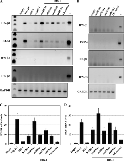
Sensory neurons latently infected with bovine herpesvirus 1 (BHV-1) abundantly express latency-related (LR) RNA (LR-RNA). Genetic evidence indicates that LR protein expression plays a role in the latency-reactivation cycle, because an LR mutant virus that contains three stop codons downstream of the first open reading frame (ORF2) does not reactivate from latency. The LR mutant virus induces higher levels of apoptotic neurons in trigeminal ganglia, and ORF2 interferes with apoptosis. Although ORF2 is important for the latency-reactivation cycle, other factors encoded by the LR gene are believed to play a supportive role. For example, two microRNAs (miRNAs) encoded within the LR gene are expressed in trigeminal ganglia of latently infected calves. These miRNAs interfere with bICP0 protein expression and productive infection in transient-transfection assays. In this report, we provide evidence that the two LR miRNAs cooperate with poly(I·C), interferon (IFN) regulatory factor 3 (IRF3), or IRF7 to stimulate beta interferon (IFN-β) promoter activity. Both miRNAs also stimulated IFN-β promoter activity and nuclear factor-kappa B (NF-κB)-dependent transcription when cotransfected with a plasmid expressing retinoic acid-inducible gene I (RIG-I). In the presence of RIG-I, the LR miRNAs enhanced survival of mouse neuroblastoma cells, which correlated with activation of the antiapoptosis cellular transcription factor, NF-κB. Immunoprecipitation assays demonstrated that both miRNAs stably interact with RIG-I, suggesting that this interaction directly stimulates the RIG-I signaling pathway. In summary, the results of these studies suggest that interactions between LR miRNAs and RIG-I promote the establishment and maintenance of latency by enhancing survival of infected neurons.


Figures

Fig 1
Fig 1
The LR mutant virus induces higher levels of IFN-β than wild-type (wt) BHV-1 in low-passage-number bovine testicle cells (BTest). BTest cells (3 × 10⁶) were mock infected (lane M) or infected with the LR mutant virus (A) or wt BHV-1 (B) at an MOI of 5. Where indicated, cultures were treated with 100 μg/ml of CHX. Cells were harvested, and total RNA was extracted at the indicated times (in hours) after infection. cDNA synthesis was subsequently performed by RT-PCR. The resultant cDNA was used as a template for PCR with primers specific for each of the three bovine IFN-β genes, as previously described (59). Genomic DNA extracted from bovine kidney cells (CRIB) served as template for the positive-control PCR (+). GAPDH cDNA amplification served as a loading control. The relative RNA levels were quantified using a biomolecular imager (Bio-Rad), and density values were normalized to a GAPDH control. The mock-infection value was set as 1. The values represent averages of the results of three independent experiments.
Fig 2
Fig 2
Schematic of LR gene and locations of sncRNAs and miRNAs. (A) Locations of the LR-RNA and bICP0 genes in the context of the BHV-1 genome. The bICP0 gene, but not the intact LR gene, is present in both repeats. The unique short (US) and unique long (UL) regions of the genome are denoted. (B) A partial restriction map of the LR gene is shown. The numbering system of the LR gene was derived from a previous study (49). The locations of the LR-RNA in cultured cells and TG are denoted relative to the bICP0 stop codon and 3′ terminus of bICP0 mRNA. The positions where the LR miRNA1 and miRNA2 are proposed to base pair with bICP0 mRNA are denoted by the black circles and were previously determined (34). The numbers in parentheses represent the BHV-1 genomic locations (GenBank accession number AJ004801). (C) Two families of LR sncRNAs were identified (34): LR sncRNA1 and LR sncRNA2. All of the sncRNA1 clones begin at position 525 of the LR gene sequence. The 5′ terminus of the sncRNA2 family begins at position 591 of the LR gene sequence. The 3′ terminus of the respective sncRNAs is variable. The positions and nucleotide sequences of the respective miRNAs within the sncRNAs are also denoted.
Fig 3
Fig 3
LR gene-encoded sncRNAs and miRNAs enhance IFN-β promoter activity stimulated by poly(I·C), IRF7, or IRF3. (A and B) BTest cells (A) or 293 cells (B) (3 × 10⁶) were cotransfected with 1 μg of CAT reporter plasmids regulated by the IFN-β1 or IFN-β3 promoter, 2 μg of pSilencer expressing the LR sncRNAs (sncRNA1 or -2), or miRNAs (miRNA1 or -2). IRF3 (1 μg), IRF7 (1 μg), or synthetic dsRNA poly(I·C) (0.5 μg) served as a positive control for the indicated cell lines and IFN-β promoters. Plasmid DNA was maintained at the same concentration by including an empty expression vector. Two days after transfection, cells were harvested and analyzed for CAT expression. The results presented represent the fold increase relative to cells transfected with the IFN-β CAT reporter plasmids plus empty pSilencer (Empty), whose value was arbitrarily set as 1. (C and D) BTest cells (C) or 293 cells (D) (3 × 10⁶) were cotransfected with 1 μg of IFN-β1 or IFN-β3 CAT plasmids, factors known to stimulate the IFN-β promoter [IRF3, IRF7 or poly(I·C)], 1 or 2 μg of LR-encoded sncRNA (sncRNA1 or -2), and 1 or 2 μg of miRNA (miRNA1 or -2). Two days after transfection, cells were harvested, and CAT activity was measured. The results shown represent the fold increase relative to the value obtained using IRF3-, IRF7-, or poly(I·C)-induced IFN-β promoter activity, whose value was arbitrarily set as 1.
Fig 4
Fig 4
The LR sncRNAs or miRNAs induced IFN-β promoter activity in BTest cells when cotransfected with RIG-I. BTest cells (3 × 10⁶) were transfected with CAT plasmids containing IFN-β1 (A) (1 μg) or IFN-β3 promoter (B) (1 μg), RIG-I (1 μg), and 2 μg of a plasmid expressing the sncRNAs (sncRNA1 or -2) or miRNAs (miRNA1 or -2). Poly(I·C) (0.5 μg) and N-RIG-I (1 μg) served as positive controls. A pSilencer control construct containing an LR gene sequence adjacent to the regions encoding the LR sncRNAs was used as a negative control (LR control; see Fig. 2C). Equivalent amounts of DNA were used for transfection by addition of an empty expression vector. Two days after transfection, cells were harvested and CAT activity was measured. The results are shown as fold increases relative to cells transfected with the IFN-β CAT reporter plasmid plus the empty pSilencer plasmid (Empty), whose value was arbitrarily set as 1. The data shown represent averages of the results of three independent experiments.
Fig 5
Fig 5
The LR sncRNAs or miRNAs induced IFN-β promoter activity in 293 cells when cotransfected with RIG-I. Human 293 cells (3 × 10⁶) were transfected with CAT plasmids containing IFN-β1 (A) (1 μg) or IFN-β3 promoter (B) (1 μg), RIG-I (1 μg), and 2 μg of a plasmid expressing the sncRNAs (sncRNA1 or -2) or miRNAs (miRNA1 or -2). Poly(I·C) (0.5 μg) and N-RIG-I (1 μg) served as positive controls. A pSilencer control construct containing an LR gene sequence adjacent to the regions encoding the small and microRNAs was used as a negative control (LR control). Equivalent amounts of DNA were used for transfection by addition of an empty expression vector. Two days after transfection, cells were harvested and CAT activity was measured. The results shown represent fold increases relative to cells transfected with the IFN-β CAT reporter plasmid plus the empty pSilencer plasmid (Empty), whose value was arbitrarily set as 1. The data shown represent averages of the results of three independent experiments.
Fig 6
Fig 6
LR-encoded sncRNAs and miRNAs increase bovine IFN-β1 and ISG54 mRNA levels when cotransfected with RIG-I. (A and B) BTest cells were transfected with the LR sncRNAs and miRNAs (2 μg) along with 1 μg of a RIG-I expression vector (A) or an empty vector (B). Poly(I·C) (0.5 μg) and N-RIG-I (1 μg) served as positive controls, and the LR control plasmid (LR control) was used as a negative control. Forty hours after transfection, cells were harvested and total RNA was extracted. cDNA synthesis was subsequently performed by RT-PCR. The resultant cDNA was used as a template for PCR with primers specific for the three bovine IFN-β genes or ISG54, as previously described (59). Genomic DNA extracted from bovine CRIB cells served as a template for the positive-control PCR (+). GAPDH amplification was used as a loading control. These results are representative of at least three independent studies. (C and D) The IFN-β1 (C) and ISG54 (D) RNA levels in agarose gels were examined using a Bio-Rad Molecular Imager FX (Molecular Dynamics, Sunnyvale, CA). The values shown are expressed as fold increases relative to the band from mock-infected cells, whose value was arbitrarily set as 1.
Fig 7
Fig 7
LR miRNAs stimulate NF-κB-dependent transcription when cotransfected with RIG-I. Approximately 2 × 10⁶ 293 cells were transfected with the 5× NF-κB luciferase reporter construct (1 μg), pRL-TK (0.033 μg), RIG-I (1 μg), 2 μg of an empty vector, the indicated LR miRNAs, or the LR control. N-RIG-I (1 μg) and poly(I·C) (0.5 μg) served as positive controls. Cells were harvested at 40 h after transfection, and the dual-luciferase assay was performed. The data represent the relative firefly luciferase activity normalized to Renilla luciferase activity. The values are expressed as fold differences relative to the pSilencer empty vector (Empty), whose value was arbitrarily set as 1.
Fig 8
Fig 8
LR miRNAs interfere with cold shock-induced apoptosis. (A) Neuro-2A cells were transfected with a plasmid encoding the β-galactosidase (β-Gal) gene (0.025 μg) and 3 μg of the empty pSilencer. Images of cells before and after cold shock-induced apoptosis are shown. Cold shock-induced apoptosis was performed as described in Materials and Methods. (B) Top panels: Neuro-2A cells were cotransfected with a plasmid encoding the β-galactosidase (β-Gal) gene (0.025 μg), 1 μg of empty pSilencer, and 2 μg of pSilencer encoding the LR miRNAs (miRNA1 or -2) or the RIG-I expression plasmid, as indicated. Bottom panels: Neuro-2A cells were cotransfected with a plasmid encoding the β-galactosidase (β-Gal) gene (0.025 μg), RIG-I (1 μg), and 2 μg of pSilencer encoding the LR miRNAs (miRNA1 or -2) or LR control, as indicated. At 48 h after transfection, Neuro-2A cells were submitted to cold shock-induced apoptosis and subsequently recovered for 3.5 h at 37°C. Cold shock-induced apoptosis was performed, and pictures of the cultures were taken. (C) The number of β-Gal-positive cells was determined, and cell survival was compared to that seen with the empty vector, whose value was arbitrarily set at 1. The values represent averages of the results of three independent studies.
Fig 9
Fig 9
The LR-encoded miRNAs interact with full-length RIG-I. (A) Schematic of experimental steps used to immunoprecipitate (IP) and amplify RIG-I-associated RNAs. (B) Monolayers containing 4 × 10⁶ 293 cells were transfected with 5 μg of an empty FLAG vector (Empty) or FLAG vectors expressing RIG-I or N-RIG-I along with 5 μg of plasmids expressing the LR miRNA1 or miRNA2 or LR control plasmid. Forty-eight hours after transfection, cells were harvested in hypotonic buffer and the RIG-I IP was performed using the FLAG monoclonal antibody. The efficiency of the IP was monitored by Western blot (WB) analysis. (C) RNA associated to the RIG-I complex was extracted by the acid-phenol:chloroform method and subsequently converted into cDNA by RT-PCR. The resultant cDNA served as a template for amplification by the reverse primers specific for the LR miRNA1 or miRNA2 or the LR control and the forward primer specific for the 5′ adaptor. These results are representative of 2 independent experiments. The first lane (M) corresponds to molecular weight markers.

Similar articles

Cited by

References

    1. Alexopoulou L, Holt AC, Medzhitov R, Flavell RA. 2001. Recognition of double-stranded RNA and activation of NF-kappaB by Toll-like receptor 3. Nature 413: 732–738 - PubMed
    1. Allen SJ, et al. 2011. The role of LAT in increased CD8+ T cell exhaustion in trigeminal ganglia of mice latently infected with herpes simplex virus 1. J. Virol. 85: 4184–4197 - PMC - PubMed
    1. Baum A, Sachidanandam R, Garcia-Sastre A. 2010. Preference of RIG-I for short viral RNA molecules in infected cells revealed by next-generation sequencing. Proc. Natl. Acad. Sci. U. S. A. 107: 16303–16308 - PMC - PubMed
    1. Bauman Y, et al. 2011. An identical miRNA of the human JC and BK polyoma viruses targets the stress-induced ligand ULBP3 to escape immune elimination. Cell Host Microbe 9: 93–102 - PubMed
    1. Belardo G, Santoro MG. 2010. Heat stress triggers apoptosis by impairing NF-kappaB sirvival signaling in malignant B cells. Leukemia 24: 187–196 - PubMed

Publication types

LinkOut - more resources