. 2012 Feb;33(5):1618-26.
doi: 10.1016/j.biomaterials.2011.11.002. Epub 2011 Nov 29.

Multifunctional, multichannel bridges that deliver neurotrophin encoding lentivirus for regeneration following spinal cord injury

Affiliations

Multifunctional, multichannel bridges that deliver neurotrophin encoding lentivirus for regeneration following spinal cord injury

Hannah M Tuinstra et al. Biomaterials. 2012 Feb.

Abstract

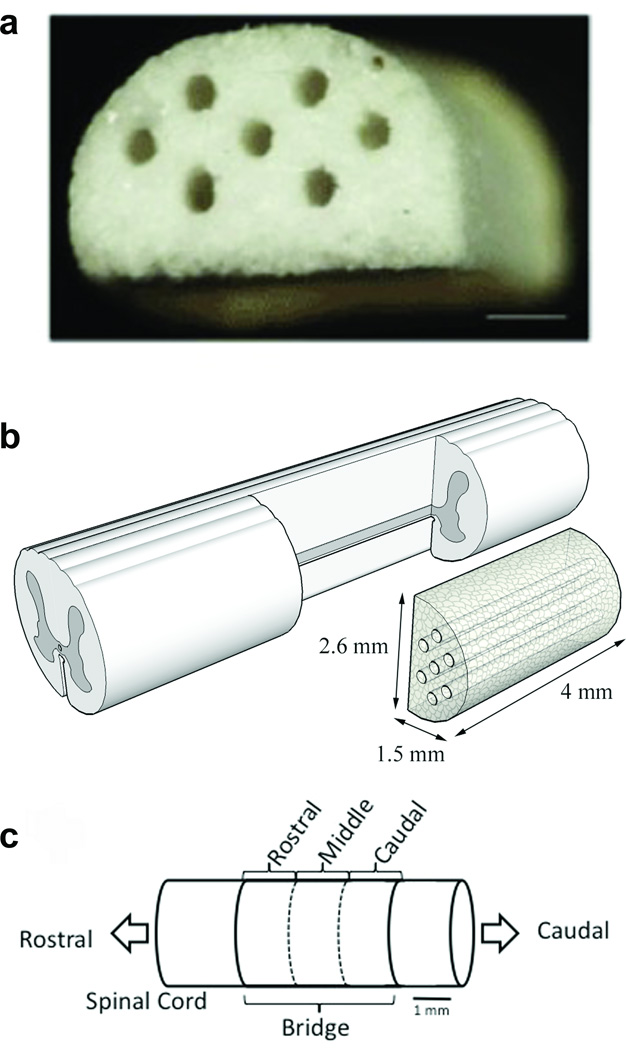
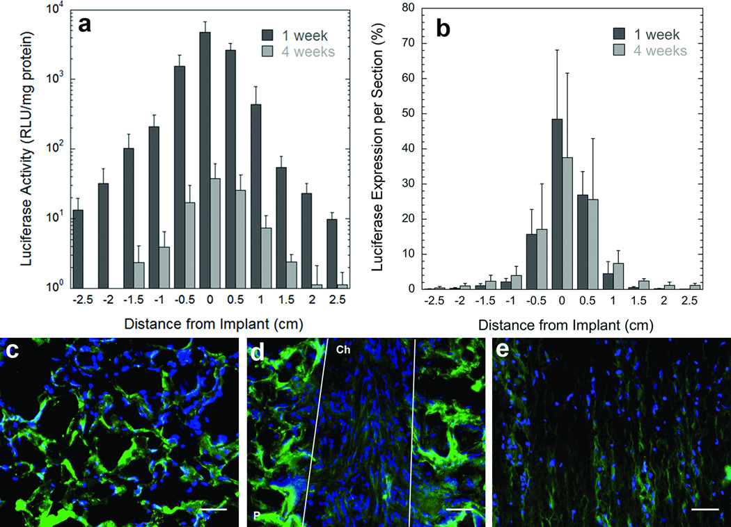
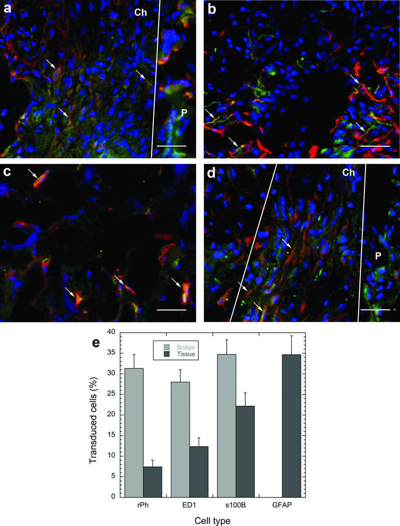
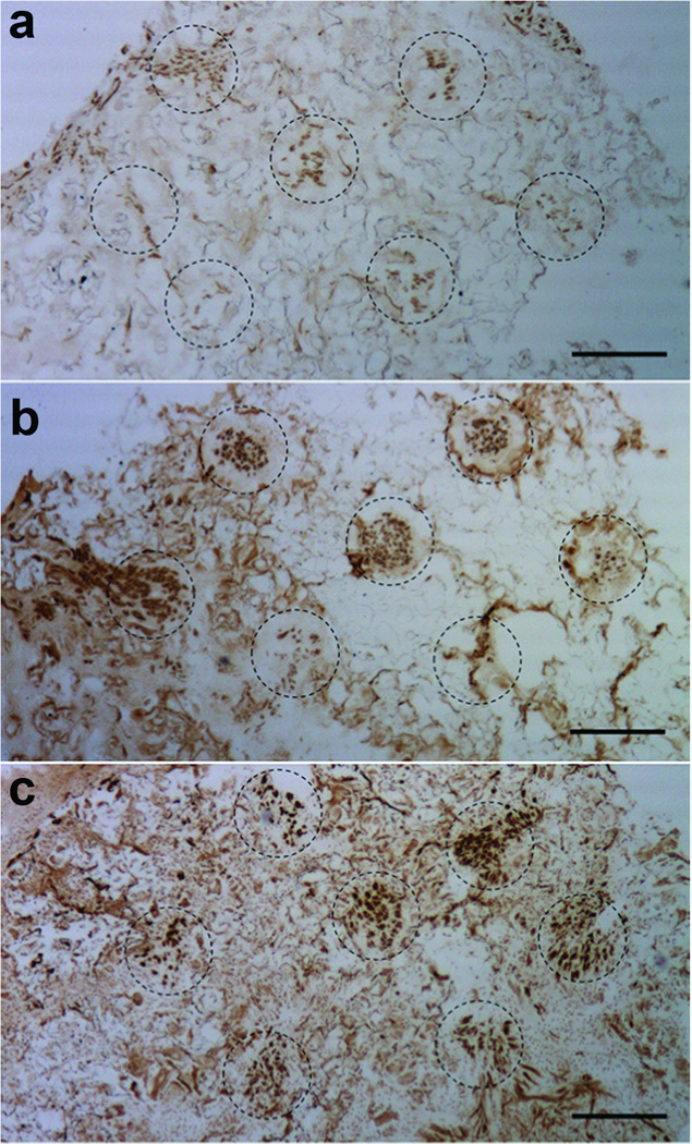
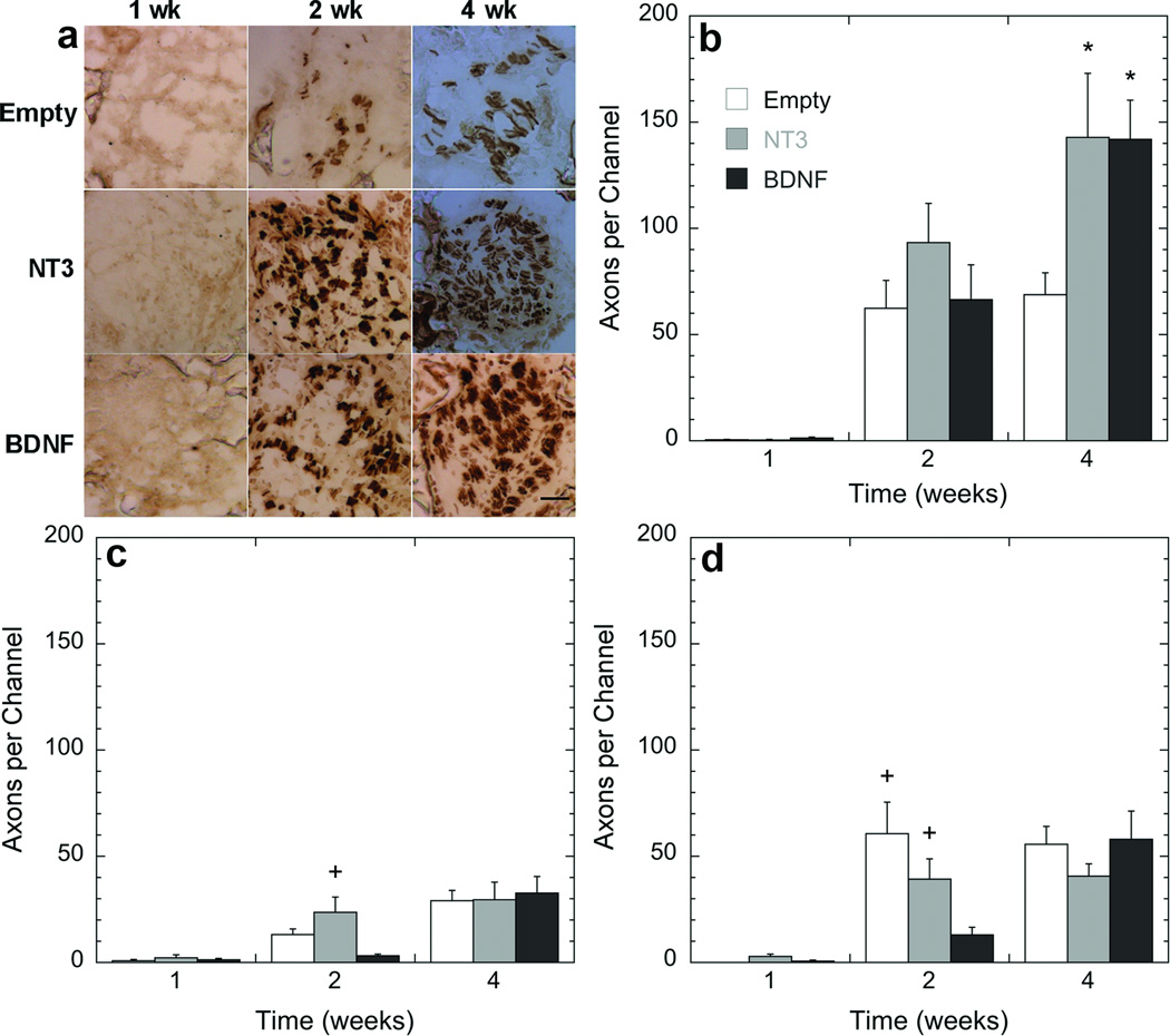
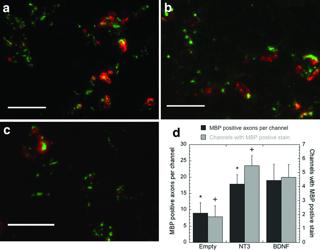
Therapeutic strategies following spinal cord injury must address the multiple barriers that limit regeneration. Multiple channel bridges have been developed that stabilize the injury following implantation and provide physical guidance for regenerating axons. These bridges have now been employed as a vehicle for localized delivery of lentivirus. Implantation of lentivirus loaded multiple channel bridges produced transgene expression that persisted for at least 4 weeks. Expression was maximal at the implant at the earliest time point, and decreased with increasing time of implantation, as well as rostral and caudal to the bridge. Immunohistochemical staining indicated transduction of macrophages, Schwann cells, fibroblasts, and astrocytes within the bridge and adjacent tissue. Subsequently, the delivery of lentivirus encoding the neurotrophic factors NT-3 or BDNF significantly increased the extent of axonal growth into the bridge relative to empty scaffolds. In addition to promoting axon growth, the induced expression of neurotrophic factors led to myelination of axons within the channels of the bridge, where the number of myelinated axons was significantly enhanced relative to control. Combining gene delivery with biomaterials to provide physical guidance and create a permissive environment can provide a platform to enhance axonal growth and promote regeneration.


Figures

Figure 1
Figure 1. Multiple channel bridges for spinal cord regeneration
(a) Photomicrograph of a multiple channel bridge showing 7 channels, each 250 µm in diameter. Scale bar is 500 µm. (b) Schematic of PLG bridge implantation in a spinal cord hemisection model. (c) Schematic of the regions in which the bridge was divided for analysis. Rostral analysis was done at 300 µm, middle at 2000 µm, and caudal at 3500 µm from the rostral edge of the bridge/tissue boundary.
Figure 2
Figure 2. Transgene expression within the spinal cord
(a) Luciferase expression in the rat spinal cord lateral hemisection model at two time points (1 week, and 4 weeks) (n=4) as a function of distance from the injury site. 0 = the bridge segment. RLU, relative light unit. (b) Percentage of total luciferase expression at each segment in the spinal cord at two time points (1 week, and 4 weeks) (n=4) as a function of distance from the injury site. 0 = the bridge segment. (c-e) Location of transduced cells after implantation of bridges loaded with a lentivirus encoding for enhanced green fluorescent protein (green). (c) Transduced cells were observed within the pores of the bridge, (d) aligned within the channels of the bridge, (e) and in the tissue adjacent to the bridge. Cell nuclei were stained with Hoechst (blue). P, polymer, Ch, channel. White lines indentify the boundaries of a channel. Scale bar is 50 µm. Error bars represent SEs.
Figure 3
Figure 3. Identification of transduced cells
Double staining of slides with antibodies to EGFP (green) and to specific cell types (red). The cell types antibodies were (a) S100β for Schwann cells, (b) GFAP for reactive astrocytes, (c) ED-1 for macrophages, or (d) rPH for fibroblasts. Cell nuclei were stained with Hoechst (blue). Arrows indicate the position of cells that stained positive for both EGFP and the specific cell antibody. P, polymer. Ch, channel. White lines indentify the boundaries of channels. Scale bar is 50 µm. Quantification of the profile of transduced cells within the bridge and the adjacent tissue (e). Error bars represent SEs.
Figure 4
Figure 4. Axon regeneration inside the channels for different treatment conditions
Images of an (a) empty bridge or a bridge loaded with a lentivirus encoding either (b) NT3 or (c) BDNF. Bridges were retrieved 4 weeks post implantation, and sections from the rostral position were stained with NF200 (neurofilament, brown). Channels are identified by the dash circles. Scale bar is 250 µm.
Figure 5
Figure 5. Quantification of axonal regeneration as a function of time and position within the bridges
(a) Time course of a single channel at the rostral position. (b-d) Axons were quantified by counting the number of NF200 positive neurofilaments inside the channels. Slides analyzed were selected from the (b) rostral (300 µm), (c) middle (2000 µm), and (d) caudal (3500 µm) regions of the bridge. Statistical analysis was done by an ANOVA with Tukey post hoc test with a p<0.05 found to be significant differently. *, significant difference compared to empty at the same time point; +, significant difference compared to BDNF at the same time point). Scale bar in a is 50 µm.
Figure 6
Figure 6. Axon myelination inside the channels
Tissue sections were stained for myelin basic protein (MBP, red) and neurofilament (NF200, green) (a-c) 4 weeks after implantation. The images are from the rostral sections of (a) a bridge loaded with a lentivirus encoding for BDNF, (b) a bridge loaded with a lentivirus encoding for NT3, and (c) an empty bridge. Scale bar is 50 µm in a-c. (d) Quantification of the MBP stain as a function of the treatment at 4 weeks post implantation and in the rostral position. Quantification was done by counting the number of myelinated axons inside the channels (■, left axis), or by counting the number of channels with positive stained myelinated axons (□, right axis). * Indicates significant difference compared to empty (p<0.01), using the Kruskal-Wallis Test. + Indicates significant difference compared to empty (p<0.01), using an ANOVA with Tukey post hoc test.

Similar articles

Cited by

References

    1. David S, Aguayo AJ. Axonal elongation into peripheral nervous system "bridges" after central nervous system injury in adult rats. Science. 1981;214:931–933. - PubMed
    1. Xu XM, Guenard V, Kleitman N, Bunge MB. Axonal regeneration into Schwann cell-seeded guidance channels grafted into transected adult rat spinal cord. J Comp Neurol. 1995;351:145–160. - PubMed
    1. Geller HM, Fawcett JW. Building a bridge: engineering spinal cord repair. Exp Neurol. 2002;174:125–136. - PubMed
    1. De Laporte L, Yang Y, Zelivyanskaya ML, Cummings BJ, Anderson AJ, Shea LD. Plasmid releasing multiple channel bridges for transgene expression after spinal cord injury. Mol Ther. 2009;17:318–326. - PMC - PubMed
    1. Yang Y, De Laporte L, Zelivyanskaya ML, Whittlesey KJ, Anderson AJ, Cummings BJ, et al. Multiple channel bridges for spinal cord injury: cellular characterization of host response. Tissue Eng Part A. 2009;15:3283–3295. - PMC - PubMed

Publication types

MeSH terms