

Isolated extradural tuberculous granuloma of the cervical spine: a case report
- PMID: 22130618
- PMCID: PMC3369063
- DOI: 10.1007/s00586-011-2095-9
Isolated extradural tuberculous granuloma of the cervical spine: a case report
Abstract
Introduction: Isolated intraspinal extradural tuberculous granuloma (IETG) without radiological evidence of vertebral involvement is uncommon, especially rare in cervical spine.
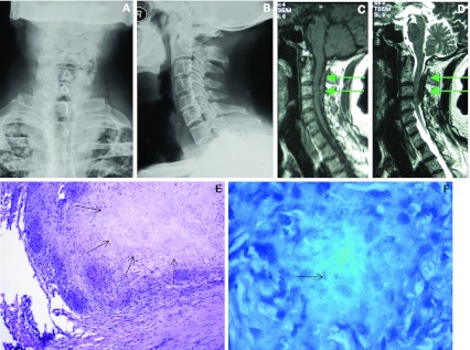
Materials and methods: We report a case of cervical IETG without bone involvement in a patient with neurological deficit. The patient suffered from progressive neurological dysfunction. MRI of cervical spine revealed an intraspinal extradural mass, and the spinal cord was edematous because of the compression. Thus C2-C4 laminectomy was performed and extradural mass was excised.
Results: The excised extradural mass was confirmed to be tuberculous granuloma through pathologic examination. Antituberculous drugs were administrated with a regular follow-up. Excellent clinical outcomes were achieved.
Conclusions: The isolated IETG, although a rare entity, should be considered in the differential diagnosis of the intraspinal mass, especially in patients with spinal cord compression and a history of tuberculosis. If there is a progressing neurological deficit, a combination of surgical and anti-tuberculous treatment should be the optimal choice.
Figures
Similar articles
-
Tuberculosis of the lower cervical spine (C5-C6) in a 24-month-old infant.Spine J. 2013 Aug;13(8):e17-20. doi: 10.1016/j.spinee.2013.02.035. Epub 2013 Mar 27. Spine J. 2013. PMID: 23541450
-
Treatment of intraspinal tuberculoma.Clin Orthop Relat Res. 2007 Jul;460:62-6. doi: 10.1097/BLO.0b013e318065b73c. Clin Orthop Relat Res. 2007. PMID: 17452917
-
Ganglion cyst of the cervical spine presenting with Brown-Sequard syndrome.J Clin Neurosci. 2006 Dec;13(10):1041-5. doi: 10.1016/j.jocn.2005.07.028. J Clin Neurosci. 2006. PMID: 17113987
-
Spinal extradural cysticercosis: a case report.Spinal Cord. 1998 Apr;36(4):285-7. doi: 10.1038/sj.sc.3100524. Spinal Cord. 1998. PMID: 9589530 Review.
-
Cervical tuberculosis associated with cervical pain and neurologic deficit: a case report and literature review.Spine J. 2014 May 1;14(5):e13-8. doi: 10.1016/j.spinee.2013.10.055. Epub 2013 Nov 19. Spine J. 2014. PMID: 24262857 Review.
Cited by
-
Non-osseous intradural tuberculoma of the thoracic spine with compressive myelopathy.Clin Case Rep. 2023 Nov 2;11(11):e8131. doi: 10.1002/ccr3.8131. eCollection 2023 Nov. Clin Case Rep. 2023. PMID: 37927983 Free PMC article.
-
Stabilizing the unstable: Tuberculosis of the odontoid process with atlanto-occipital instability-Case report and review of literature.Clin Case Rep. 2023 Dec 28;12(1):e8379. doi: 10.1002/ccr3.8379. eCollection 2024 Jan. Clin Case Rep. 2023. PMID: 38161635 Free PMC article.
-
Long-Segment Ventral Spinal Epidural Abscesses Caused by Mycobacterium Tuberculosis: A Report of 2 Cases and Review.JB JS Open Access. 2025 Jun 20;10(2):e24.00249. doi: 10.2106/JBJS.OA.24.00249. eCollection 2025 Apr-Jun. JB JS Open Access. 2025. PMID: 40547101 Free PMC article. Review.
-
Concurrent Spinal Epidural Tubercular and Pyogenic Abscess of Cervical Spine without Bony Involvement.J Neurosci Rural Pract. 2019 Apr-Jun;10(2):374-378. doi: 10.4103/jnrp.jnrp_318_18. J Neurosci Rural Pract. 2019. PMID: 31001041 Free PMC article.
References
-
- Davidson PT, Horowitz I (1970) Skeletal tuberculosis—A review with patient presentations and discussion. Am J Med 48:77 - PubMed
-
- Hsu LCS, Leong JCY. Tuberculosis of the lower cervical-spine (C2–C7)—A report on 40 cases. J Bone Joint Surg Br. 1984;66:1–5. - PubMed
-
- Arseni C, Samitca DCT (1960) Intraspinal tuberculous granuloma. Brain 83:285 - PubMed
Publication types
MeSH terms
LinkOut - more resources
Full Text Sources
Medical
Miscellaneous
