Review
. 2011 Dec;17(6):659-76.
doi: 10.1177/1073858411386615.

Viruses and multiple sclerosis

Affiliations
Review

Viruses and multiple sclerosis

Gregory P Owens et al. Neuroscientist. 2011 Dec.

Abstract

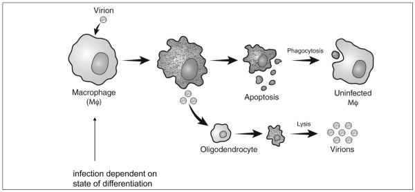
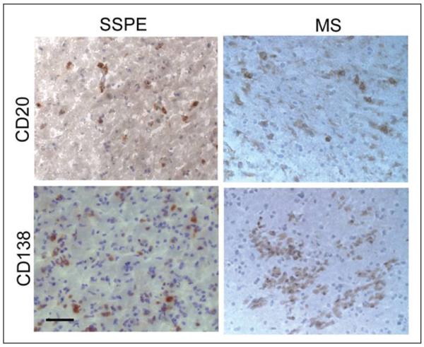
Multiple sclerosis (MS) is a chronic demyelinating disorder of unknown etiology, possibly caused by a virus or virus-triggered immunopathology. The virus might reactivate after years of latency and lyse oligodendrocytes, as in progressive multifocal leukoencephalopathy, or initiate immunopathological demyelination, as in animals infected with Theiler's murine encephalomyelitis virus or coronaviruses. The argument for a viral cause of MS is supported by epidemiological analyses and studies of MS in identical twins, indicating that disease is acquired. However, the most important evidence is the presence of bands of oligoclonal IgG (OCBs) in MS brain and CSF that persist throughout the lifetime of the patient. OCBs are found almost exclusively in infectious CNS disorders, and antigenic targets of OCBs represent the agent that causes disease. Here, the authors review past attempts to identify an infectious agent in MS brain cells and discuss the promise of using recombinant antibodies generated from clonally expanded plasma cells in brain and CSF to identify disease-relevant antigens. They show how this strategy has been used successfully to analyze antigen specificity in subacute sclerosing panencephalitis, a chronic encephalitis caused by measles virus, and in neuromyelitis optica, a chronic autoimmune demyelinating disease produced by antibodies directed against the aquaporin-4 water channel.


Figures

Figure 1
Figure 1
Direct immunofluorescence with a 1 to 20 dilution of antibody to human IgG conjugated to fluorescein isothiocyanate (green fluorescence) shows IgG deposition at the junction of plaque-periplaque white matter (middle arrow), in mononuclear cells (bottom arrow), and in normal white matter (top arrow). The antigen against which the IgG in the multiple sclerosis brain and CSF is directed is unknown.
Figure 2
Figure 2
Proposed model of Theiler’s virus persistence in macrophages leading to demyelination.
Figure 3
Figure 3
CD20+ B cells and CD138+ plasma cells in subacute sclerosing panencephalitis (SSPE) and multiple sclerosis (MS) brain. Frozen sections of SSPE or MS brain were stained with antibodies to CD20 or CD138 antigen (brown) and counterstained with hematoxylin. Bar = 50μm.
Figure 4
Figure 4
Histopathologic sections of spinal cord from a myelin basic protein-induced experimental allergic encephalomyelitis rat transfused with rat AQP4-specific CSF rAb 10 (Bennett and others 2009). H & E = hematoxylin/eosin staining reveals perivascular vacuolization and mononuclear and polymorphonuclear infiltrates (arrowheads). LFB = Luxol fast blue/PAS staining demonstrates perivascular demyelination and PAS+ macrophages (asterisks). C9neo = perivascular deposition of C9neo indicates local complement activation (arrowheads). GFAP = glial fibrillary acidic protein reveals extensive perivascular astrocyte loss associated with demyelination (asterisk). hIgG, human IgG deposition surrounding regions of perivascular demyelination (arrowheads). APP = amyloid precursor protein staining shows axonal transections (arrowheads).
Figure 5
Figure 5
VH family gene segment use in multiple sclerosis (MS) CSF plasma blasts differ significantly from use in peripheral blood CD19+ B lymphocytes.
Figure 6
Figure 6
Recombinant antibodies derived from a mouse anti-MOG hybridoma stain MOG-transfected HEK 293 cells. MOG = myelin-oligodendrocyte protein.
Figure 7
Figure 7
Panning phage-displayed random peptide libraries with antibody-coated wells. Three major steps include binding of the libraries with target antibody, washing away nonspecific phage, and eluting bound phage with a low pH buffer. Three to five rounds are performed to enrich specific phage peptides.
Figure 8
Figure 8
Streamlined protocol to determine specificity of phage peptides. Individual phage plaque from second and third pans amplified in U96 DeepWell plates are first screened for positivity against panning antibody using enzyme-linked immunosorbent assay (ELISA). Potential positive clones were confirmed by duplicate ELISA with preimmune human IgG/irrelevant rAb as negative control, followed by phage DNA purification and sequence analysis.

References

    1. Ascherio A, Munch M. Epstein-Barr virus and multiple sclerosis. Epidemiology. 2000;11:220–4. - PubMed
    1. Ascherio A, Munger KL, Lennette ET, Spiegelman D, Hernan MA, Olek MJ. Epstein-Barr virus antibodies and risk of multiple sclerosis: a prospec-tive study. JAMA. 2001;286:3083–8. others. - PubMed
    1. Baranzini SE, Jeong MC, Butunoi C, Murray RS, Bernard CC, Oksenberg JR. B cell repertoire diversity and clonal expansion in multiple sclerosis brain lesions. J Immunol. 1999;163:5133–44. - PubMed
    1. Baranzini SE, Mudge J, van Velkinburgh JC, Khankhanian P, Khrebtukova I, Farmer N. Genome, epigenome and RNA sequences of monozygotic twins discordant for multiple sclerosis. Nature. 2010;464:1351–6. others. - PMC - PubMed
    1. Barnett MH, Prineas JW. Relapsing and remitting multiple sclerosis: pathology of the newly forming lesion. Ann Neurol. 2004;55:458–68. - PubMed

Publication types

MeSH terms