

Localization of pain-related brain activation: a meta-analysis of neuroimaging data
- PMID: 22131304
- PMCID: PMC6869965
- DOI: 10.1002/hbm.21416
Localization of pain-related brain activation: a meta-analysis of neuroimaging data
Abstract
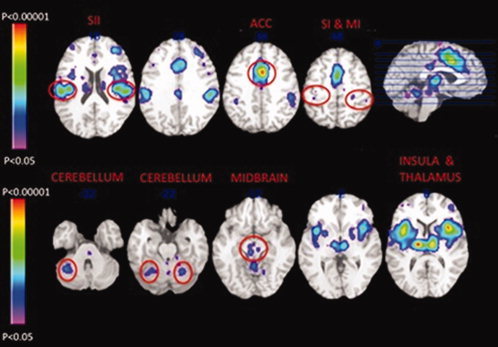
A meta-analysis of 140 neuroimaging studies was performed using the activation-likelihood-estimate (ALE) method to explore the location and extent of activation in the brain in response to noxious stimuli in healthy volunteers. The first analysis involved the creation of a likelihood map illustrating brain activation common across studies using noxious stimuli. The left thalamus, right anterior cingulate cortex (ACC), bilateral anterior insulae, and left dorsal posterior insula had the highest likelihood of being activated. The second analysis contrasted noxious cold with noxious heat stimulation and revealed higher likelihood of activation to noxious cold in the subgenual ACC and the amygdala. The third analysis assessed the implications of using either a warm stimulus or a resting baseline as the control condition to reveal activation attributed to noxious heat. Comparing noxious heat to warm stimulation led to peak ALE values that were restricted to cortical regions with known nociceptive input. The fourth analysis tested for a hemispheric dominance in pain processing and showed the importance of the right hemisphere, with the strongest ALE peaks and clusters found in the right insula and ACC. The fifth analysis compared noxious muscle with cutaneous stimuli and the former type was more likely to evoke activation in the posterior and anterior cingulate cortices, precuneus, dorsolateral prefrontal cortex, and cerebellum. In general, results indicate that some brain regions such as the thalamus, insula and ACC have a significant likelihood of activation regardless of the type of noxious stimuli, while other brain regions show a stimulus-specific likelihood of being activated.
Copyright © 2011 Wiley Periodicals, Inc.
Figures
References
-
- Adler LJ, Gyulai FE, Diehl DJ, Mintun MA, Winter PM, Firestone LL ( 1997): Regional brain activity changes associated with fentanyl analgesia elucidated by positron emission tomography. Anesth Analg 84: 120–126. - PubMed
-
- Aharon I, Becerra L, Chabris CF, Borsook D ( 2006): Noxious heat induces fMRI activation in two anatomically distinct clusters within the nucleus accumbens. Neurosci Lett 392: 159–164. - PubMed
-
- Amano N, Hu JW, Sessle BJ ( 1986): Responses of neurons in feline trigeminal subnucleus caudalis (medullary dorsal horn): To cutaneous, intraoral, and muscle afferent stimuli. J Neurophysiol 55: 227–243. - PubMed
-
- Andersson JL, Lilja A, Hartvig P, Langstrom B, Gordh T, Handwerker H, Torebjork E ( 1997): Somatotopic organization along the central sulcus, for pain localization in humans, as revealed by positron emission tomography. Exp Brain Res 117: 192–199. - PubMed
Publication types
MeSH terms
Substances
Grants and funding
LinkOut - more resources
Full Text Sources
Medical
