. 2011 Nov 30;31(48):17480-7.
doi: 10.1523/JNEUROSCI.2262-11.2011.

Getting drowsy? Alert/nonalert transitions and visual thalamocortical network dynamics

Affiliations

Getting drowsy? Alert/nonalert transitions and visual thalamocortical network dynamics

Yulia Bereshpolova et al. J Neurosci. .

Abstract

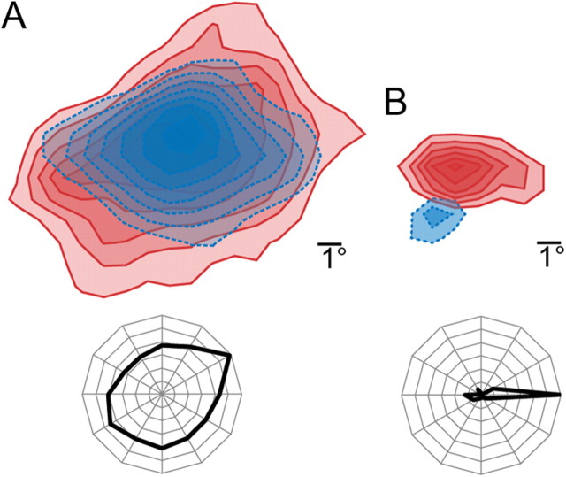
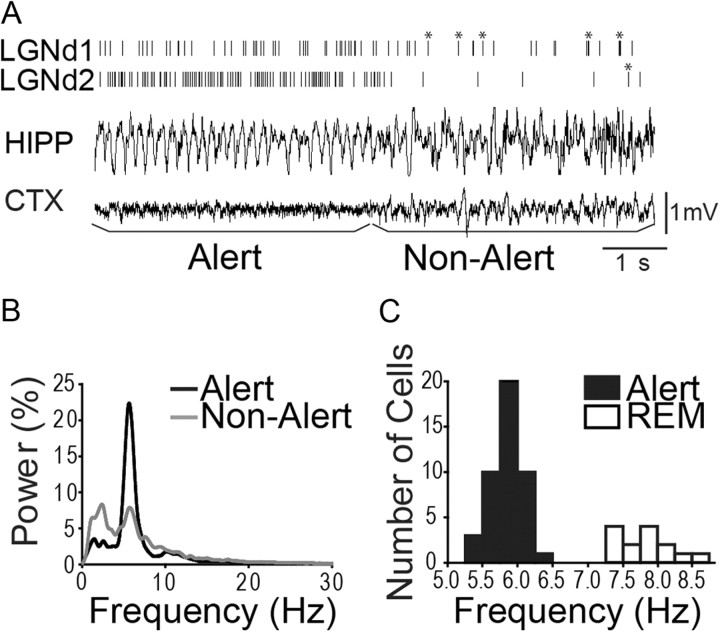
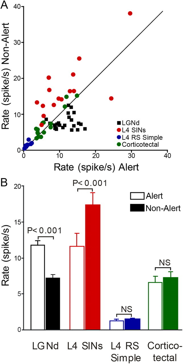
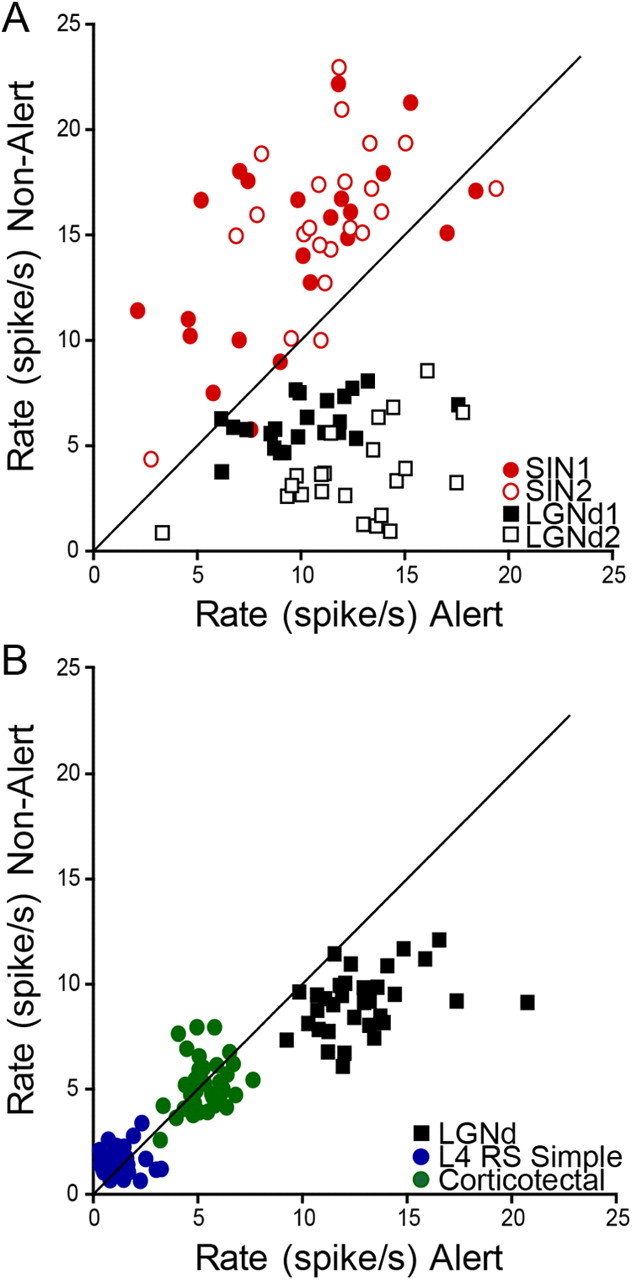
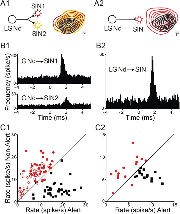
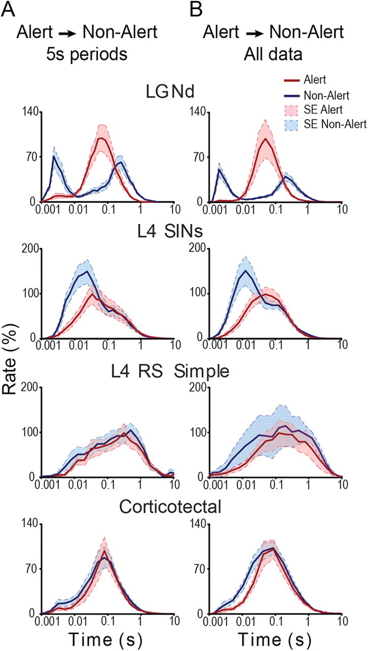
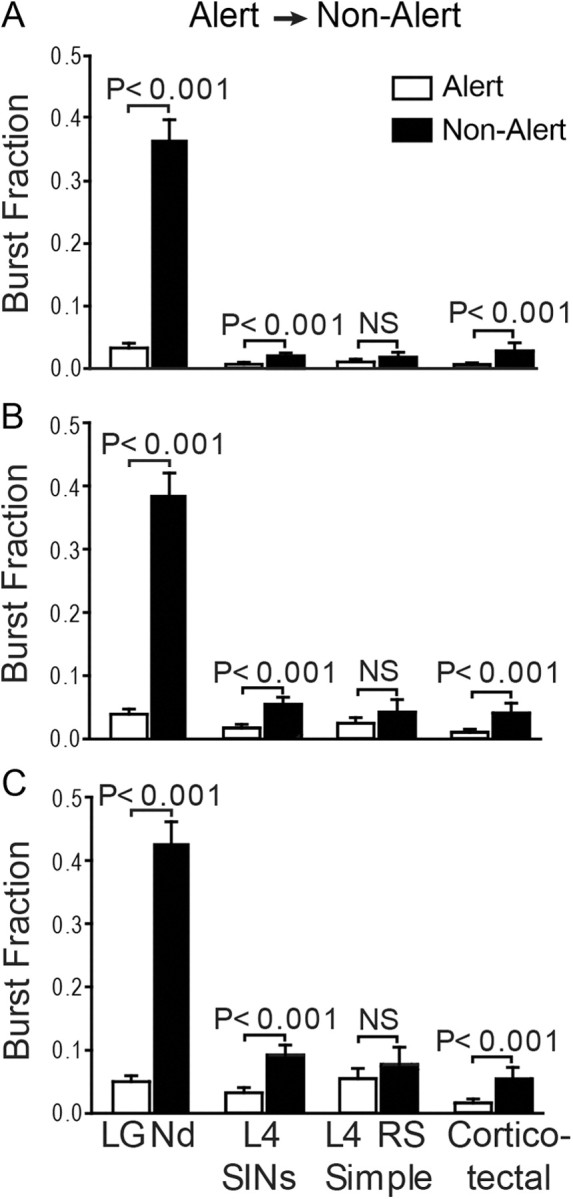
The effects of different EEG brain states on spontaneous firing of cortical populations are not well understood. Such state shifts may occur frequently under natural conditions, and baseline firing patterns can impact neural coding (e.g., signal-to-noise ratios, sparseness of coding). Here, we examine the effects of spontaneous transitions from alert to nonalert awake EEG states in the rabbit visual cortex (5 s before and after the state-shifts). In layer 4, we examined putative spiny neurons and fast-spike GABAergic interneurons; in layer 5, we examined corticotectal neurons. We also examined the behavior of retinotopically aligned dorsal lateral geniculate nucleus (LGNd) neurons, usually recorded simultaneously with the above cortical populations. Despite markedly reduced firing and sharply increased bursting in the LGNd neurons following the transition to the nonalert state, little change occurred in the spiny neurons of layer 4. However, fast-spike neurons of layer 4 showed a paradoxical increase in firing rates as thalamic drive decreased in the nonalert state, even though some of these cells received potent monosynaptic input from the same LGNd neurons whose rates were reduced. The firing rates of corticotectal neurons of layer 5, similarly to spiny cells of layer 4, were not state-dependent, but these cells did become more bursty in the nonalert state, as did the fast-spike cells. These results show that spontaneous firing rates of midlayer spiny populations are remarkably conserved following the shift from alert to nonalert states, despite marked reductions in excitatory thalamic drive and increased activity in local fast-spike inhibitory interneurons.


Figures

Figure 1.
Figure 1.
Receptive fields of putative spiny neurons and SINs of layer 4. A, An example of the receptive field of SIN, constructed using the reverse correlation method from response to sparse noise stimulation. The receptive field of SIN has spatially overlapping ON (red) and OFF (blue) subregions (top). In contrast to RS simple cells, SINs are either not sensitive to orientation or exhibit only a weak orientation bias (bottom). B, The receptive field of a layer 4 RS simple cell has a strong ON (red) subregion and a small and weaker OFF (blue) region (top). These cells show high orientation selectivity to drifting gratings (bottom).
Figure 2.
Figure 2.
Spontaneous firing rates and burst rates of thalamic neurons change dramatically between alert and nonalert states. A, An example of the transition from an alert to nonalert state, as defined by hippocampal (HIPP) EEG. Cortical (CTX) EEG is shown below. Vertical lines in top two traces indicate spike trains from two simultaneously recorded LGNd cells, with bursts marked by asterisks. B, The power spectral density functions of hippocampal EEG during alert (black line) and nonalert (gray line) periods for the two LGNd neurons shown in A. These LGNd cells underwent 36 transitions from alert to nonalert states, and the FFTs were calculated by first summing all the 5 s alert periods and then summing all the 5 s nonalert periods. The y-axis shows the percentage of power contained within each bin of 0.5 Hz. C, Filled bars, Distribution of the peak frequency in power spectral density functions during the alert state for all of neurons we studied. These frequencies clearly contrast with those recorded during periods for REM sleep (open bars).
Figure 3.
Figure 3.
Spontaneous firing rates of LGNd and cortical neurons during alert and nonalert states. A, Each data point represents the mean firing rate across all state transitions of a single neuron of the different classes studied in both alert and nonalert states. B, The population average of spontaneous firing rates for each class of neurons studied during different states of vigilance.
Figure 4.
Figure 4.
Within-cell consistency of responses to state transitions. A, Relation between state and spontaneous firing rate in simultaneously recorded neurons studied during repeated transitions from alert to nonalert state. Two LGNd cells (black open and filled squares) and two SINs (red open and filled circles) underwent 22 shifts from alert to nonalert states, and each data point represents the spontaneous activity during the 5 s before and 5 s after each of these shifts. B, Another example, where simultaneous recordings were obtained from an LGNd neuron (black squares), a corticotectal neuron (green circles) and a layer 4 RS simple cell (blue circles). These cells underwent 34 state shifts from alert to nonalert states.
Figure 5.
Figure 5.
State-dependent opposing shifts in firing frequency between synaptically connected SINs and LGNd neurons. A1, B1, C1, Spontaneous action potentials of a single LGNd neuron were simultaneously recorded with those of two retinotopically aligned cortical SINs. A1, The proposed circuit and the retinotopic alignment of LGNd and SIN receptive fields. B1, Cross-correlograms of the spike trains of the thalamocortical neuron with the two SINs. C1, Scatter plots for each of these three neurons during the 5 s before and after alert to nonalert shifts. SINs are shown as open and filled circles, LGNd neurons as black squares. A2, B2, C2, Same as A1, B1, and C1, respectively, but for another LGNd neuron and a monosynaptically connected V1 SIN.
Figure 6.
Figure 6.
The state-related distribution of interspike intervals in different cell types. A, The ISI histograms of different cell classes during alert (red lines) and nonalert (blue lines) epochs. Only the 5 s periods before and after the transitions from alert to nonalert state are included, and data from all of the cells of each class (normalized) are included. The bimodal shape of the distribution for LGNd cells and SINs reflects the intervals occurring within and between bursts. Alert and nonalert states are shown by solid and dashed lines, respectively. B, The same analysis as in A, but composed of a much larger dataset that included all alert and nonalert segments during the entire data file. The shaded areas in A and B show the SEM periods.
Figure 7.
Figure 7.
A–C, Rapid changes in bursting of LGNd neurons and some cortical populations within 5 s of a state switch from alert to nonalert. Bars indicate percentage of spikes (±SE) that are part of the bursts for the different cell classes during alert (open) and nonalert (filled) epochs. Bursts were defined as two or more spikes with a preceding interval >100 ms and an ISI of either ≤4 (A), ≤7 (B), or ≤10 (C) ms. LGNd cells, SINs, and corticotectal neurons, but not RS simple cells, showed increased bursting in the nonalert state.

References

    1. Ahmed B, Anderson JC, Douglas RJ, Martin KA, Nelson JC. Polyneuronal innervation of spiny stellate neurons in cat visual cortex. J Comp Neurol. 1994;341:39–49. - PubMed
    1. Alonso JM, Usrey WM, Reid RC. Precisely correlated firing in cells of the lateral geniculate nucleus. Nature. 1996;383:815–819. - PubMed
    1. Bereshpolova Y, Amitai Y, Gusev AG, Stoelzel CR, Swadlow HA. Dendritic backpropagation and the state of the awake neocortex. J Neurosci. 2007;27:9392–9399. - PMC - PubMed
    1. Bezdudnaya T, Cano M, Bereshpolova Y, Stoelzel CR, Alonso JM, Swadlow HA. Thalamic burst mode and inattention in the awake LGNd. Neuron. 2006;49:421–432. - PubMed
    1. Buzsaki G, Bickford RG, Ponomareff G, Thal LJ, Mandel R, Gage FH. Nucleus basalis and thalamic control of neocortical activity in the freely moving rat. J Neurosci. 1988;8:4007–4026. - PMC - PubMed

Publication types

LinkOut - more resources