. 2011 Nov 30;31(48):17612-21.
doi: 10.1523/JNEUROSCI.3449-11.2011.

CCAAT/enhancer binding protein-δ expression by dendritic cells regulates CNS autoimmune inflammatory disease

Affiliations

CCAAT/enhancer binding protein-δ expression by dendritic cells regulates CNS autoimmune inflammatory disease

Vicky W W Tsai et al. J Neurosci. .

Abstract

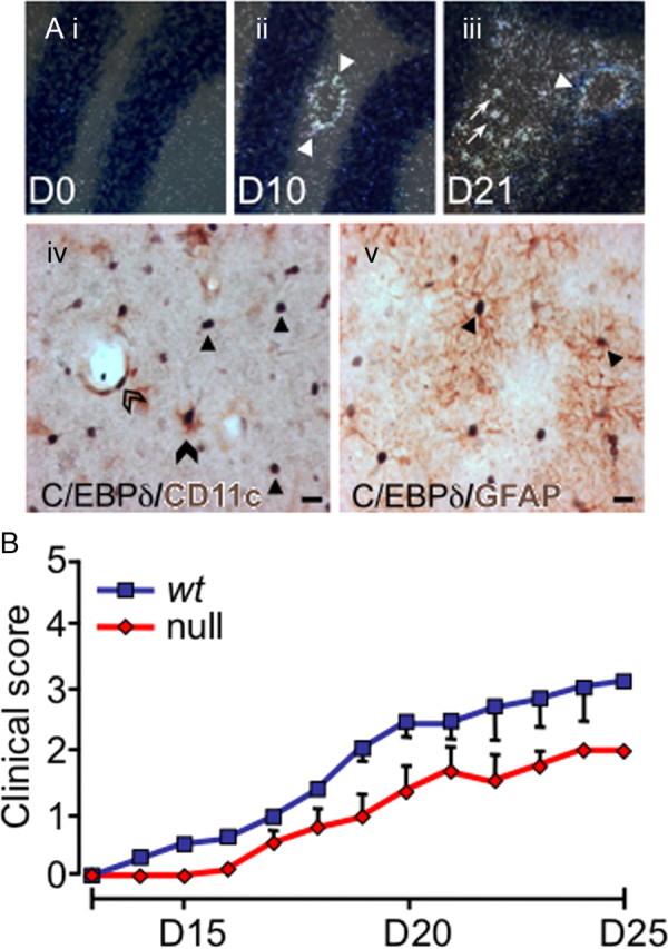
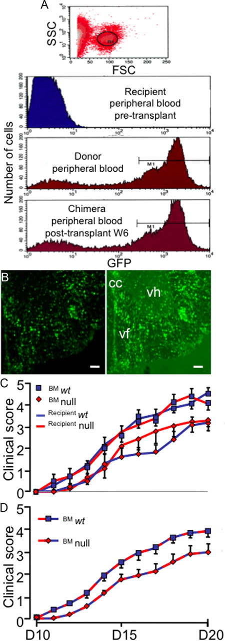
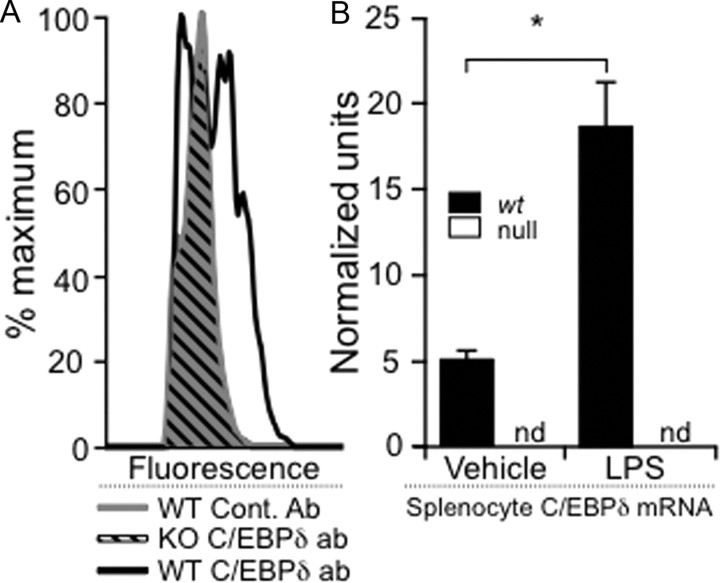
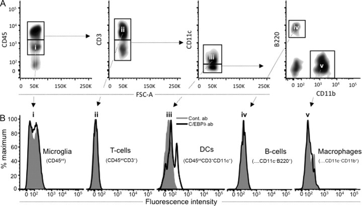
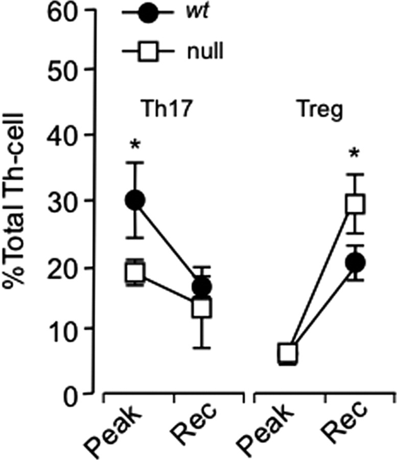
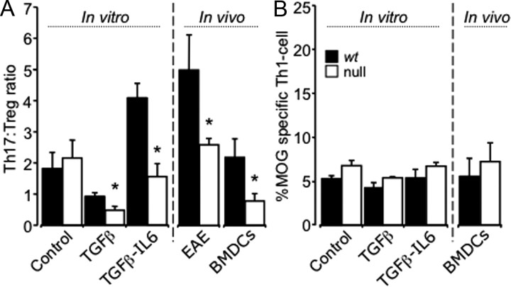
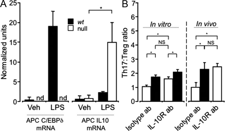
CCAAT enhancer binding protein-delta (C/EBPδ) is a transcription factor that regulates inflammatory processes mediating bystander neuronal injury and CNS autoimmune inflammatory disease. The mechanism of the involvement of C/EBPδ in these processes remains to be determined. Here, we examined the cellular source(s) and mechanisms by which C/EBPδ may be involved in an animal model of multiple sclerosis. Mice deficient in C/EBPδ expression exhibited less severe clinical disease than wild-type littermates in response to induction of experimental autoimmune encephalomyelitis (EAE) by vaccination with a myelin oligodendrocyte glycoprotein (MOG) fragment. This reduction in EAE severity was associated with a significant alteration in the complement of major CNS T-helper (Th) cell subtypes throughout disease, manifest as reduced ratios of Th17 cells to regulatory T-cells (Tregs). Studies in bone marrow chimeric mice indicated that C/EBPδ expression by peripherally derived immune cells mediates C/EBPδ involvement in EAE. Follow up in vitro and in vivo examination of dendritic cell (DC) mediated Th-cell development suggests that C/EBPδ suppresses DC expression of interleukin-10 (IL-10), favoring Th17 over Treg development. In vitro and in vivo blockade of IL-10 signaling attenuated the effect of reduced C/EBPδ expression by DCs on Th17:Treg ratios. These findings identify C/EBPδ as an important DC transcription factor in CNS autoimmune inflammatory disease by virtue of its capacity to alter the Th17:Treg balance in an IL-10 dependent fashion.


Figures

Figure 1.
Figure 1.
C/EBPδ is expressed centrally in EAE, and EAE severity is reduced in C/EBPδ-null mutants. Ai–iii, C/EBPδ is upregulated throughout EAE. Wild-type animals were killed at day 10 (D10; preclinical EAE), and day 21 (D21; peak disease), and compared with unvaccinated (day 0) animals (n = 3/group). Representative sections of the cerebellum are shown. Before vaccination (D0), C/EBPδ mRNA is not detectable. At D10, C/EBPδ is expressed in cells associated with the cerebral vasculature (arrowheads). C/EBPδ is further upregulated in the perivascular region (arrowhead) and is present in parenchymal cells (arrows) at day 21 (D21). iv, Dual immunohistochemical labeling demonstrates that C/EBPδ protein is expressed in DCs and astrocytes in EAE. Dual staining for CD11c (brown) and C/EBPδ (black) was performed on spinal cord sections from D21 EAE mice. DCs expressing nuclear C/EBPδ (black) and cytoplasmic/surface CD11c (brown) are seen in the CNS parenchyma (filled arrowhead) and perivascular (open arrowhead) regions. Many C/EBPδ-positive nuclei are not associated with CD11c coexpression (arrows). Substituting anti-CD11c for anti-MHC class II gave a similar pattern of single and dual staining (data not shown). v, Dual labeling for the astrocyte-specific marker GFAP (brown) and C/EBPδ (black) identify remaining C/EBPδ+CD11c cells as astrocytes (arrows). Scale bars, 10 μm. B, EAE severity is reduced in C/EBPδ-deficient mice. Animals were vaccinated with MOG35–55 and clinical scores monitored daily. The clinical severity of disease was reduced by one full grade in C/EBPδ−/− (n = 6 per group; p = 0.019; repeated-measures ANOVA). Error bars represent SEM.
Figure 2.
Figure 2.
Absent C/EBPδ expression in peripheral immune cells is responsible for reduced EAE in null mice. A, Six weeks after irradiation and BMT, most circulating cells are of donor origin. BM cells from ubiquitous GFP-expressing animals were transferred to irradiated wt mice. High forward scatter cells were gated and assessed for GFP expression (R1). This was undetectable in peripheral blood of recipients before transplantation, while most peripheral blood mononuclear cells of donor animals expressed GFP. Six weeks (W6) after BMT the profile of mononuclear cell GFP expression in recipient mice was similar to that of donors (n = 6). B, Fluorescence image (right) of ventral spinal cord from a mouse at day 21 (D21) after EAE induction (clinical score = 3) showing infiltration of transplanted GFP cells into the cord. Infiltrating cells displayed morphological features of both lymphocytes and APCs. Left, Infiltrating, bone-marrow derived, GFP-fluorescent cells. Right, Fluorescence overlaid with dark field to provide anatomical detail (cc, central canal; vf, ventral funiculus; vh, ventral horn). Scale bars, 100 μm. C, Chimeric animals were generated and EAE induced 6 weeks later and animals clinically scored as a measure of disease. There was no significant difference in disease between wt and C/EBPδ−/− (null) animals when circulating mononuclear cells were C/EBPδ+/+ (n = 9 per group; p = 0.54; pooled from 2 independent experiments). Similarly, there was no difference in EAE severity when wt and C/EBPδ−/− hosts had circulating mononuclear cells that were C/EBPδ−/− (n = 9 per group; p = 0.25; pooled from 2 independent experiments). D, Reduced C/EBPδ expression in circulating mononuclear cells is responsible for reduced EAE in C/EBPδ−/− animals. As the differences in EAE severity appeared dependent on the genotype of circulating mononuclear cells, data were pooled based on the genotype of donor bone marrow. Animals whose circulating monocytes were C/EBPδ−/− displayed a significant reduction in EAE disease (n = 18 per group; p = 0.0003; repeated-measures ANOVA). All error bars represent SEM.
Figure 3.
Figure 3.
Validation of an intracellular staining method for detecting C/EBPδ in bone marrow-derived mononuclear cells. A, C/EBPδ was detected by intracellular protein labeling. To validate an intracellular FACS staining method, we demonstrated that intracellular C/EBPδ labeling was not present in C/EBPδ−/− splenocytes treated with LPS (diagonal black lines), which could not be discriminated from similarly treated wt splenocytes labeled with control antibody (solid gray). However, wt splenocytes treated with LPS clearly demonstrated C/EBPδ labeling (black outline) compared with these controls. B, To confirm that LPS treatment was likely to lead to increased C/EBPδ protein, mRNA expression was assayed 4 h after activation of splenocytes with LPS. Quantitative rtPCR demonstrated significant upregulation of C/EBPδ mRNA in wt splenocytes compared with vehicle-treated cells (black). C/EBPδ mRNA could not be amplified (nd, not detected) from C/EBPδ−/− (null) splenocytes (white). *p < 0.05.
Figure 4.
Figure 4.
Dendritic cells are the only CNS APCs expressing C/EBPδ protein in EAE. CNS mononuclear cells were isolated from EAE animals on day 10, before disease onset. C/EBPδ-specific labeling was demonstrated in CD3CD11c+ cells indicating a DC phenotype. A, CD45+ cell subsets of the CNS, including microglia, T-cells, B-cells, DCs, and macrophages, were identified by gating on surface markers CD45, CD3, B220, CD11c, and CD11b (Bailey et al., 2007). B, C/EBPδ protein expression was not detected in microglia that were identified on the basis of their intermediate CD45 expression (i; CD45int). Of the cells expressing higher levels of CD45 (CD45hi), only DCs (iii; CD45hiCD3CD11c+) were found to express C/EBPδ protein, while T-cells (ii; CD45hiCD3+), B-cells (iv; CD45hiCD3CD11cB220+CD11b), and macrophages (v; CD45hiCD3CD11cB220CD11b+) did not.
Figure 5.
Figure 5.
C/EBPδ−/− animals have fewer Th17 and more Treg cells in the CNS over the course of EAE. wt and C/EBPδ−/− (null) mice were vaccinated with MOG, and CNS mononuclear cells were isolated near the peak of disease (day 18; n = 8) and at partial recovery (day 25; n = 16). CNS mononuclear cells were surface stained with anti-CD45, anti-CD3, and anti-CD4 and then labeled for intracellular FOXP3, IL-17, and INFγ. Data represent the pooled results of two independent experiments. The proportions of Th17 to Treg cells in wt mice were comparable to those reported in murine EAE (Korn et al., 2007). C/EBPδ−/− animals had fewer Th17 cells at peak disease and more Tregs during the recovery phase of EAE (Rec), with the resultant Th17:Treg ratios being significantly reduced at both time points (p = 0.02, p = 0.03). Error bars represent SEM. *p < 0.05.
Figure 6.
Figure 6.
Expression of C/EBPδ by CD11c+ DCs directs Th17/Treg cell differentiation in vitro and in vivo. A, 2D2 transgenic mice that received intracerebral injections of BMDCs from wt and C/EBPδ−/− (null) BM had similar numbers of CD3+, CD4+, and Vβ11+ Th-cells recruited to the CNS (p = 0.58). Matured, MOG antigen-pulsed BMDCs were cocultured with MOG-reactive wt 2D2 Th-cells (CD3+CD4+) under Treg and Th17 polarizing conditions. Under vehicle control conditions there was no significant difference in the development of Th17, Treg, or Th1 T-cells directed by wt versus C/EBPδ−/− BMDCs (p = 0.46). With Treg- and Th17-polarizing conditions there was a significant reduction in the Th17:Treg ratio directed by C/EBPδ−/− BMDCs (p = 0.04; p = 0.005 n = 9 per group). In EAE (Fig. 5) there was also a significantly decreased Th17:Treg ratio in vivo between wt and C/EBPδ−/− animals (p = 0.02). When BMDCs were injected into the CNS of 2D2 animals, differentiation of MOG antigen-specific Th-cells was again skewed by C/EBPδ−/− BMDCs, leading to significantly a reduced Th17:Treg ratio (p = 0.02; n = 6). B, For both in vivo and in vitro groups there were no significant differences in Th1 development when cells were activated in vitro (control, p = 0.29; TGFβ, p = 0.67, or TGFβ and IL-6, p = 0.60) or in vivo (p = 0.2). *p < 0.05; all error bars represent SEM.
Figure 7.
Figure 7.
Expression of C/EBPδ by DCs modulates Th17:Treg ratios via IL-10. A, Total mRNA was isolated 2 h after LPS or vehicle stimulation of MOG antigen-pulsed wt and C/EBPδ−/− (null) BMDCs, and qPCR assessment of IL-10 and C/EBPδ mRNA was performed. LPS treatment led to a significant increase in C/EBPδ mRNA, which was not detectible (nd) in C/EBPδ−/− BMDCs. Upon treatment with LPS, both C/EBPδ−/− and wt BDMCs significantly upregulated IL-10 mRNA (p = 0.046). However, C/EBPδ−/− BMDCs produced significantly more IL-10 mRNA than wt under the same stimulation conditions (p = 0.046). B, In vitro and in vivo experiments in Figure 6A were repeated using Th17-polarizing conditions, with, and without, IL-10 receptor blockade (IL-10R ab). In vitro incubation with isotype control antibody showed again that C/EBPδ−/− BMDCs mediated a significantly reduced Th17:Treg ratio (p = 0.005). Blockade of IL-10 signaling significantly increased the Th17:Treg ratio mediated by C/EBPδ−/− DCs (p = 0.01; n = 9), but did not completely negate the effect of C/EBPδ deficiency in DCs. wt or C/EBPδ−/− MOG antigen-pulsed, matured DCs were injected into the CNS of 2D2 mice undergoing isotype or specific IL-10R antibody treatment every other day. Blocking IL-10 signaling again lead to a significant increase in the Th17:Treg ratio generated by C/EBPδ−/− BMDCs (p = 0.012), which did not differ significantly from that induced by isotype antibody-treated wt BMDCs in vivo (p = 0.78). Data represent n = 9; NS, p > 0.2; *p < 0.05; all error bars represent SEM.

Similar articles

Cited by

References

    1. Alam T, An MR, Papaconstantinou J. Differential expression of three C/EBP isoforms in multiple tissues during the acute phase response. J Biol Chem. 1992;267:5021–5024. - PubMed
    1. Arora M, Poe SL, Oriss TB, Krishnamoorthy N, Yarlagadda M, Wenzel SE, Billiar TR, Ray A, Ray P. TLR4/MyD88-induced CD11b(+)Gr-1(int)F4/80(+) non-migratory myeloid cells suppress Th2 effector function in the lung. Mucosal Immunol. 2010;3:578–593. - PMC - PubMed
    1. Bailey SL, Schreiner B, McMahon EJ, Miller SD. CNS myeloid DCs presenting endogenous myelin peptides ‘preferentially’ polarize CD4(+) T(H)-17 cells in relapsing EAE. Nat Immunol. 2007;8:172–180. - PubMed
    1. Bailey-Bucktrout SL, Caulkins SC, Goings G, Fischer JA, Dzionek A, Miller SD. Cutting edge: central nervous system plasmacytoid dendritic cells regulate the severity of relapsing experimental autoimmune encephalomyelitis. J Immunol. 2008;180:6457–6461. - PMC - PubMed
    1. Ben-Sasson SZ, Hu-Li J, Quiel J, Cauchetaux S, Ratner M, Shapira I, Dinarello CA, Paul WE. IL-1 acts directly on CD4 T cells to enhance their antigen-driven expansion and differentiation. Proc Natl Acad Sci U S A. 2009;106:7119–7124. - PMC - PubMed

Publication types

MeSH terms

Substances

LinkOut - more resources