. 2011 Nov 30;31(48):17736-51.
doi: 10.1523/JNEUROSCI.1482-11.2011.

Congenital CNS hypomyelination in the Fig4 null mouse is rescued by neuronal expression of the PI(3,5)P(2) phosphatase Fig4

Affiliations

Congenital CNS hypomyelination in the Fig4 null mouse is rescued by neuronal expression of the PI(3,5)P(2) phosphatase Fig4

Jesse J Winters et al. J Neurosci. .

Abstract

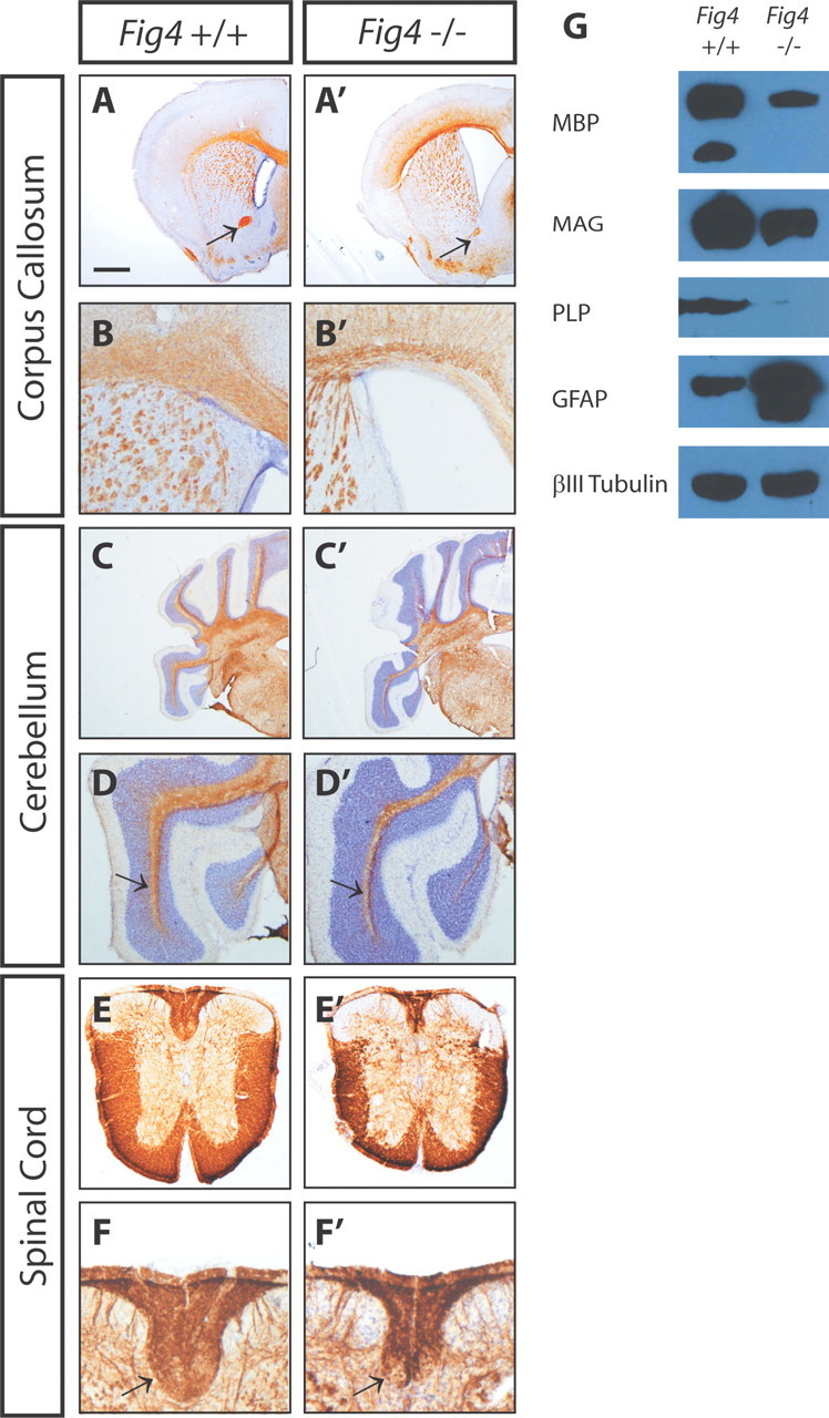
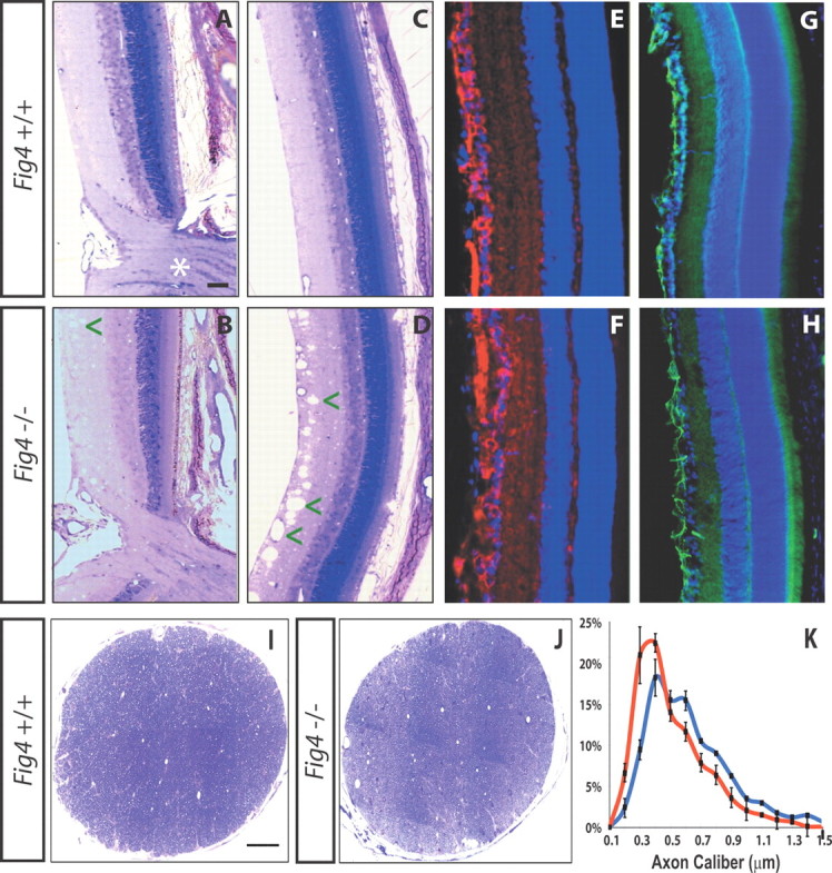
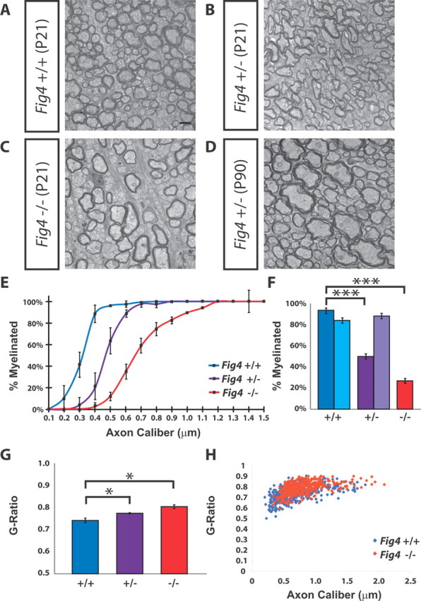
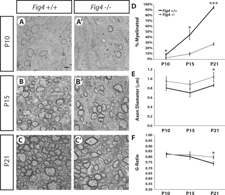
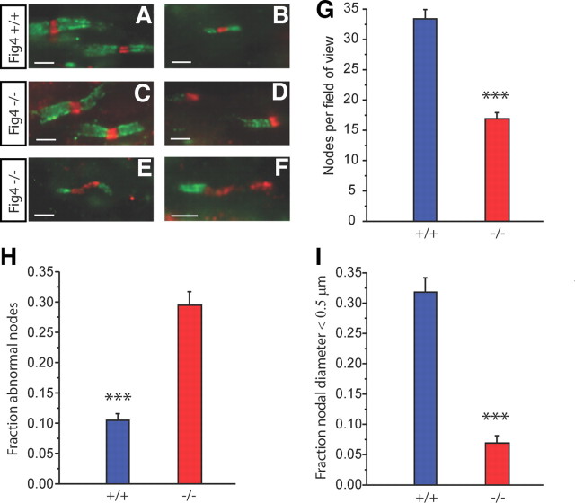
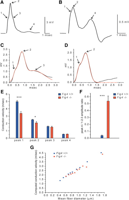
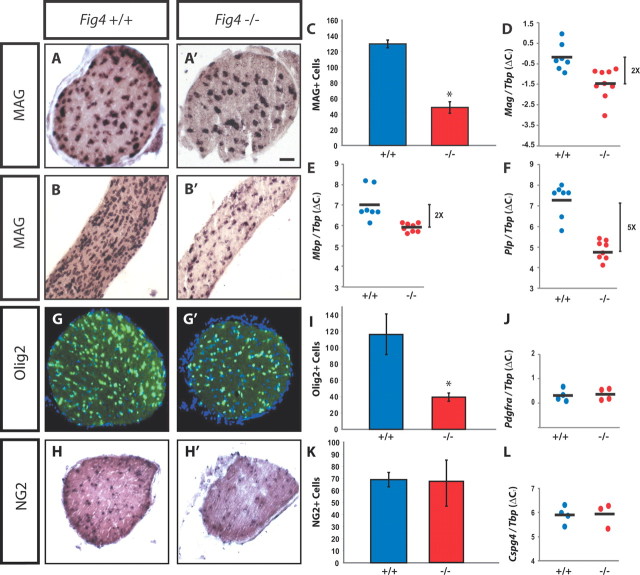
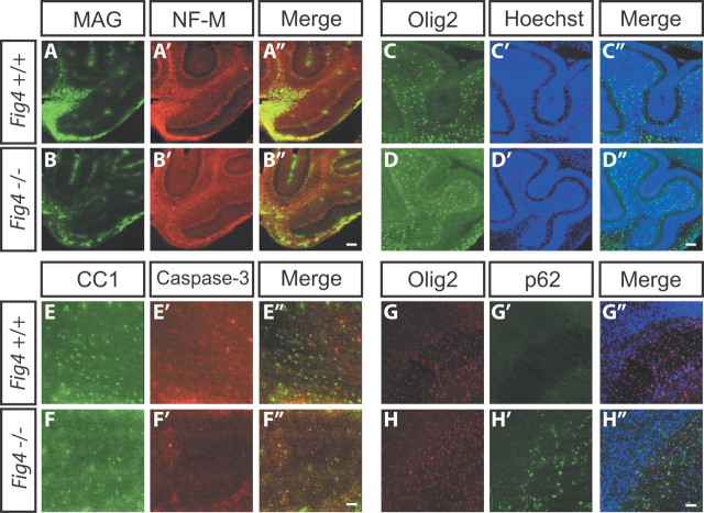
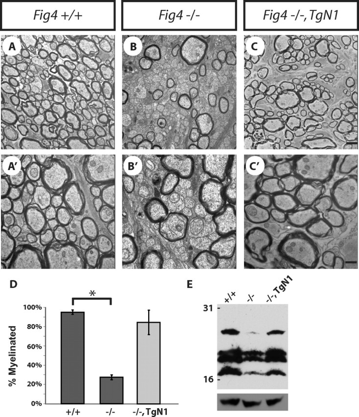
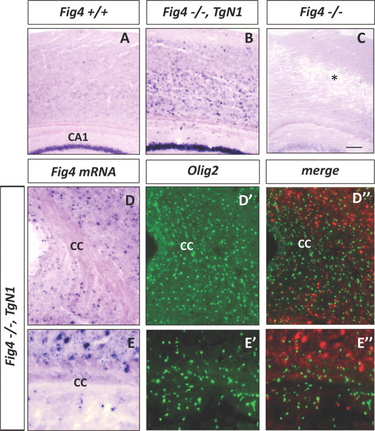
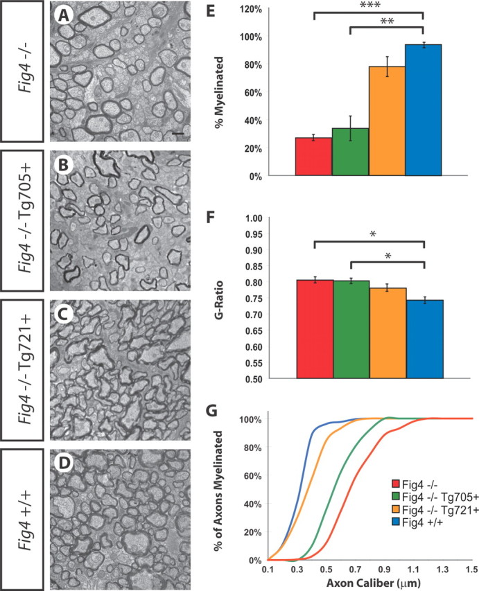
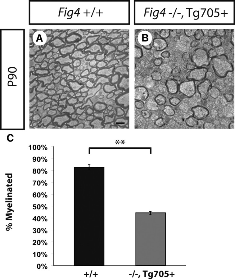
The plt (pale tremor) mouse carries a null mutation in the Fig4(Sac3) gene that results in tremor, hypopigmentation, spongiform degeneration of the brain, and juvenile lethality. FIG4 is a ubiquitously expressed phosphatidylinositol 3,5-bisphosphate phosphatase that regulates intracellular vesicle trafficking along the endosomal-lysosomal pathway. In humans, the missense mutation FIG4(I41T) combined with a FIG4 null allele causes Charcot-Marie-Tooth 4J disease, a severe form of peripheral neuropathy. Here we show that Fig4 null mice exhibit a dramatic reduction of myelin in the brain and spinal cord. In the optic nerve, smaller-caliber axons lack myelin sheaths entirely, whereas many large- and intermediate-caliber axons are myelinated but show structural defects at nodes of Ranvier, leading to delayed propagation of action potentials. In the Fig4 null brain and optic nerve, oligodendrocyte (OL) progenitor cells are present at normal abundance and distribution, but the number of myelinating OLs is greatly compromised. The total number of axons in the Fig4 null optic nerve is not reduced. Developmental studies reveal incomplete myelination rather than elevated cell death in the OL linage. Strikingly, there is rescue of CNS myelination and tremor in transgenic mice with neuron-specific expression of Fig4, demonstrating a non-cell-autonomous function of Fig4 in OL maturation and myelin development. In transgenic mice with global overexpression of the human pathogenic FIG4 variant I41T, there is rescue of the myelination defect, suggesting that the CNS of CMT4J patients may be protected from myelin deficiency by expression of the FIG4(I41T) mutant protein.


Figures

Figure 1.
Figure 1.
CNS hypomyelination in the Fig4−/− mouse. P21 WT (Fig4+/+) and mutant (Fig4−/−) brain and spinal cord sections were immunostained with anti-MBP (brown) and counterstained with Nissl (purple). A, A′, Coronal sections of forebrain show labeling of the corpus callosum and rostral branch of the anterior commissure (arrow). B, B′, Close-up of forebrain showing thinning of the corpus callosum and enlarged lateral ventricles in Fig4−/− compared with Fig4+/+ mice. C, C′, Coronal sections at the level of cerebellum and brainstem. D, D′, Close-up of cerebellum shows thinning of white matter (arrow) in lobules of Fig4−/− mice. E, E′, Cross-sections of thoracic spinal cord show loss of white matter in Fig4−/− mutants. F, F′, Close-up of dorsal funiculus shows decreased MBP labeling in Fig4−/− compared with Fig4+/+ spinal cord. Arrows point to corticospinal tract. G, Representative immunoblots of equal protein amounts of P21 Fig4+/+ and Fig4−/− brain membranes. A reduction of the myelin-associated proteins MBP, PLP, and MAG is observed in Fig4−/− brain membranes. The most pronounced reduction is observed for PLP. GFAP is greatly increased in Fig4−/− mutants, although no changes in neuron-specific class III β-tubulin are found. Scale bar: A, A′, C, C′, 1 mm; B, B′, D, D′, 0.3 mm; E, E′, 0.5 mm; F, F′, 0.2 mm.
Figure 2.
Figure 2.
No evidence for loss of RGCs or optic nerve axons in Fig4−/− mice. A–D, Toluidine blue labeling of P21 WT (Fig4+/+) and mutant (Fig4−/−) retina. No major defects in retinal lamination or the optic nerve head (asterisk) are observed in Fig4−/−mutants. In Fig4−/− mice, the inner retina shows small-diameter (arrowhead in B) and sometimes large-diameter (arrowheads in D) vacuoles. E, F, P19 retinae labeled with TuJ1 to visualize RGCs (red) and counterstained with the Hoechst nuclear dye 33342 (blue). G, H, P19 retinae labeled with anti-GFAP (green) and counterstained with a nuclear dye (blue). Scale bars: A–H, 50 μm. I, J, Low-magnification images of optic nerve cross-sections of P21 Fig4+/+ and Fig4−/− mice stained with toluidine blue. Scale bars: I, J, 50 μm. K, Morphometric assessment of axon caliber distribution in P21 optic nerve of Fig4+/+ (blue graph, n = 4) and Fig4−/− (red graph, n = 4) mice. Measurements of axon diameter were made from electron microscopy images and revealed a shift toward smaller-sized axons in Fig4−/− mutant mice.
Figure 3.
Figure 3.
Small-caliber axons are not myelinated in the Fig4−/− optic nerve. A–C, Electron microscopy images of optic nerve cross-sections of P21 Fig4+/+, Fig4+/−, and Fig4−/− mice shows a Fig4 gene dosage-dependent decrease in the percentile of myelinated axons. D, Image of optic nerve cross-section of Fig4+/− at P90. E, Graph showing the percentage of myelinated axons at P21 as a function of axon caliber within each genotype. Fifteen different size groups, ranging from 0.1 to 1.5 μm in axon caliber, were used to subdivide optic nerve axons. Three mice for each genotype (Fig4+/+, Fig4+/−, and Fig4−/−) were included in the analysis. Differences in the percentile of myelination between Fig4+/+ and Fig4−/− are not significant for axons <0.2 μm; significant for axons between 0.2 and 0.3 μm (p < 0.05), highly significant for axons between 0.3 and 1.0 μm (p < 0.007), and not significant for axons between 1.1 and 1.5 μm in diameter. Results are presented as the mean ± SEM; Student's t test. F, Quantification of percentile of myelinated axons in the optic nerve at P21 [dark blue (Fig4+/+), dark purple (Fig4+/−), and red (Fig4−/−)] and at P90 [light blue (Fig4+/+) and light purple (Fig4+/−)]. At P21, 94 ± 2% (Fig4+/+, n = 4 mice), 50 ± 2% (Fig4+/−, n = 4), and 27 ± 2% (Fig4−/−, n = 3 mice) of axons are myelinated. At P90, 84 ± 2% (Fig4+/+, n = 3 mice) and 88 ± 2% (Fig4+/−, n = 4 mice) of axons are myelinated. Results are presented as the mean ± SEM; ***p < 0.001, Student's t test. G, Quantification of g-ratio for all myelinated axons in P21 Fig4+/+, Fig4+/−, and Fig4−/− optic nerve. Results are presented as the mean ± SEM; *p < 0.05 (n = 4 mice for each genotype). H, Scatter plot showing the distribution of g-ratios of myelinated axons as a function of axon caliber at P21 for Fig4+/+ (blue) and Fig4−/− (red) optic nerves (n = 4). Scale bars: A–D, 1 μm.
Figure 4.
Figure 4.
Fig4−/− mice exhibit severe dysmyelination of the optic nerve. Developmental time study of axon myelination. Ultrastructural images of optic nerve cross-sections from Fig4+/+and Fig4−/− mice at P10 (A, A′), P15 (B, B′), and P21 (C, C′) are shown. Scale bar, 1 μm. D, Quantification of percentage of myelinated axons in Fig4+/+and Fig4−/− optic nerve at P10 (n = 3), P15 (n = 3), and P21 (n = 4). E, Quantification of average diameter of myelinated axons in Fig4+/+and Fig4−/− optic nerve at P10 (n = 3), P15 (n = 3), and P21 (n = 4). F, Fiber g-ratios at different developmental stages were determined and are shown as a function of age at P10 (n = 3), P15 (n = 3), and P21 (n = 4). Only myelinated fibers were included to determine g-ratios. D–F, Results are presented as the mean ± SEM; *p < 0.05, ***p < 0.001, Student's t test.
Figure 5.
Figure 5.
Disorganized nodes of Ranvier and paranodes in the Fig4−/− optic nerve. A–F, Optic nerve sections immunostained for Na+ channels (red) and Caspr (green) to visualize nodes and paranodes, respectively. Fig4+/+ (A, B) and Fig4−/− (C–F) optic nerve at P21. A, C, Normal nodal structure in large-diameter axons is found in both genotypes. B, A thin myelinated axon, common in Fig4+/+ but rare in Fig4−/− mice. Abnormal structure in Fig4−/− mutants included focal heminodes (D), retracted paranodes (E), and heminodes with broad regions of Na+ channel label (F). Bar graphs in G–I plot means ± SEM. G, The number of nodes per FOV (68 × 55 μm) is decreased in Fig4−/− mice. H, The fraction of nodes that fit into at least one of the categories shown in D–F is much greater in Fig4−/− mice. I, Plot of the fraction of nodes present on axons with a diameter ≤0.5 μm. FOVs analyzed: Fig4+/+, n = 29; Fig4−/−, n = 27. ***p < 0.001, Student's t test. Scale bars: A–F, 10 μm.
Figure 6.
Figure 6.
Larger population of slow conducting axons in the optic nerve of Fig4−/− mice. A, CAP recorded from a Fig4+/+optic nerve at P22–P24. Peaks 1–3 reflect contributions from myelinated axons. There is only a small signal from slow unmyelinated axons (peak 4). B, CAP recorded from an optic nerve of an age-matched Fig4−/− mouse. In contrast to Fig4+/+ optic nerve, a very large slow component (peak 4) is seen in mutants. The component likely representing thinner myelinated axons (peak 3) is small or absent in mutants. C, Peaks 1–3 of a Fig4+/+ nerve (black line) are fitted by the sum of three Gaussians (red line). D, Optic nerve from a Fig4−/− mutant in which the fit requires only peaks 1 and 2 (red line). E, Conduction velocities of peaks 1–3 (from Gaussian fits) and peak 4 in Fig4+/+ and Fig4−/− optic nerves. F, The ratio of the amplitude of peak 4 to the maximum amplitude of the myelinated complex (peaks 1–3). Fig4+/+, n = 5 animals (9 nerves), Fig4−/−, n = 5 animals (10 nerves). Error bars indicate SEM; *p < 0.05, ***p < 0.001, Student's t test. G, Predicted conduction velocity computed from measured g-ratios. g-ratio data from electron micrographs of Fig4+/+ fibers (n = 399) and Fig4−/− fibers (n = 400) were sorted by diameter into 10 groups of each genotype, and the mean values from each group were used in calculations. Action potentials were calculated at nodes 5–8 at 37°C. The graph thus represents predicted velocities based on the measured g-ratios, i.e., myelin thickness. Note that CAP velocities in Fig4−/− nerve (red dots) deviate from the linear plot and are decreased compared with WT control (blue dots). The model does not include any nodal/paranodal abnormalities.
Figure 7.
Figure 7.
Reduction of myelinating OLs in Fig4−/− mice. P21 optic nerve sections of WT (Fig4+/+) and mutant (Fig4−/−) mice were stained with markers for the OL lineage. A–B′, MAG in situ hybridization on optic nerve cross-sections and longitudinal sections. C, Quantification of MAG+ cells per optic nerve cross-sections at P21 in Fig4+/+ (n = 4) and Fig4−/− (n = 4) mice. D–F, qRT-PCR on P21 whole-brain RNA normalized to the TATA box binding protein Tbp shows a decrease in copy number of MAG (p = 0.0029), MBP (p = 0.0017), and PLP (p < 0.0001) in Fig4−/− (n = 8) compared with Fig4+/+ (n = 7) mice. G, G′, Olig2 (green) immunostaining of P21 optic nerve cross-sections counterstained with Hoechst nuclear dye 33342 (blue). I, Quantification of the mean number of Olig2+ cells in optic nerve of Fig4+/+ (116 ± 24) and Fig4−/− (40 ± 5) mice (n = 3). J–L, At P21, OPCs are not decreased in Fig4−/− optic nerve or whole brain compared with Fig4+/+ mice. J, qRT-PCR for PDGFRα of Fig4+/+ (n = 4) and Fig4−/− (n = 4) brain revealed no significant difference (p = 0.74). H, H′, NG2 in situ hybridization on P21 optic nerve cross-sections. K, Quantification of NG2+ cells per optic nerve cross-section at P21 in Fig4+/+ and Fig4−/− mice (n = 2 mice per genotype). L, qRT-PCR for NG2 (Cspg4) of Fig4+/+ (n = 4) and Fig4−/− (n = 4) of RNA isolated from 3-week-old brains revealed no significant difference (p = 0.96). Results are presented as the mean ± SEM; *p < 0.05, Student's t test. Scale bar: A, A′, 50 μm; B, B′, 100 μm; G, G′, 50 μm; H, H′, 80 μm.
Figure 8.
Figure 8.
OPCs are not significantly reduced in the Fig4−/− mice. Sagittal brain sections (25 μm) of P7 WT (Fig4+/+) and mutant (Fig4−/−) pups. A–B″, Double-immunofluorescence labeling of the cerebellum with anti-MAG (green) and anti-NF-M (red). C–D″, P7 cerebellar lobules labeled with anti-Olig2 (green) and Hoechst for nuclear staining (blue). The number of Olig2+ cells per FOV in cerebellar lobules of Fig4+/+ (44 ± 7, n = 3) is not significantly different from Fig4−/−mice (39 ± 6, n = 3). E–F″, Pons of P7 pups stained with CC1 antibody to visualize postmitotic OLs (green) and double stained with anti-activated caspase-3 (red) to assess cell death. Few cells with activated caspase-3 were detected in the pons, and these cells did not overlap with CC1+ OLs. Note that the number of CC1-labeled cells in the Fig4−/− pons is decreased by 46 ± 9% compared with age-matched WT controls. p < 0.01, Student's t test. G–H″, At P7, before the onset of forebrain myelination, the number of Olig2+ cells (red) within the corpus callosum per FOV is not significantly different between Fig4+/+ (167 ± 15, n = 3) and Fig4−/− (152 ± 10, n = 3) mice. At this age, many p62-positive cells (green) are observed in the corpus callosum of Fig4−/− but not Fig4+/+pups. Double immunofluorescence revealed no overlap between p62- and Olig2-positive cells in the corpus callosum. Sections were counterstained with Hoechst nuclear dye 33342 (blue). Cell counts are presented as the mean ± SEM. Scale bars: A–B″, C–D″, G–H″, 60 μm; E–F″, 30 μm.
Figure 9.
Figure 9.
Neuron-specific expression of Fig4 is sufficient to rescue the Fig4−/− myelination defect in vivo. Ultrastructural analysis of optic nerve cross-sections of Fig4+/+ (A, A′) and Fig4−/− (B, B′) mice and transgenic Fig4−/−,TgN1 mice (C, C′). D, Quantification of the fraction of myelinated optic nerve axons revealed greatly enhanced myelination in Fig4−/−,TgN1 mice (85 ± 12%) compared with Fig4−/− mice (27 ± 2%). The percentile of myelinated axons in the Fig4−/−,TgN1 optic nerve is similar to Fig4+/+ mice (92 ± 2%). E, Western blot analysis of whole-brain extracts of P25 Fig4+/+ (lane 1), Fig4−/− (lane 2), and Fig4−/−,TgN1 (lane 3) mice shows that neuronal expression of WT Fig4 on a Fig4 null background is sufficient to restore MBP expression to WT levels. Anti-MBP recognizes four bands between ∼17 and 27 kDa. Anti-GAPDH is shown as loading control. Results are presented as the mean ± SEM; *p < 0.01, Student's t test. Scale bar: A–C, 1 μm; A′–C′, 0.3 μm.
Figure 10.
Figure 10.
Fig4−/−,TgN1 transgenic mice express Fig4 in neurons but not OLs. A–C, Cross-sections of P21 neocortex of WT mice (Fig4+/+), NSE-transgenic mice crossed onto a Fig4−/− background (Fig4−/−,TgN1), and Fig4 null (Fig4−/−) mice were hybridized with a digoxigenin-labeled cRNA probe specific for Fig4. In Fig4+/+ sections, there is weak labeling in the neocortex and robust staining of hippocampal CA1 pyramidal neurons. In the Fig4−/−,TgN1 mice, there are many strongly labeled cells in the neocortex and intense labeling of CA1 pyramidal neurons. In Fig4−/− null mice, there is degeneration in the neocortex (asterisk) and no hybridization signal. D–D″, Low-magnification images of the corpus callosum (cc) of Fig4−/−,TgN1 brain stained for Fig4 transcript and Olig2 protein. Note in D″, showing merged Fig4 and Olig2 labeling, the Fig4 hybridization signal is shown in red. E–E″, Higher magnification of the corpus callosum demonstrates primarily non-overlapping labeling of Olig2 and Fig4 in Fig4−/−,TgN1 brain. Scale bar: A–C, 100 μm; D–D″, 64 μm; E–E″, 20 μm.
Figure 11.
Figure 11.
Dose-dependent rescue of Fig4−/− dysmyelination phenotype by overexpression of the human pathogenic variant Fig4I41T. Ultrastructural images of P21 optic nerve section of Fig4 null mice (Fig4−/−) (A), transgenic mice expressing low levels of Fig4I41T (line 705) on a Fig4 null background (Fig4−/−,Tg705) (B), transgenic mice expressing high levels of Fig4I41T (line 721) on a Fig4 null background (Fig4−/−,Tg721) (C), and WT (Fig4+/+) (D) mice. E, Quantification of percentage of myelinated axons for Fig4−/− (red, n = 4), Fig4−/−,Tg705 (green, n = 3), Fig4−/−,Tg721 (yellow, n = 4), and Fig4+/+ (blue, n = 4) optic nerve. F, g-ratio of myelinated axons in P21 Fig4−/− (n = 4), Fig4−/−,Tg705 (n = 3), Fig4−/−,Tg721 (n = 4), and Fig4−/− (n = 4) optic nerve. G, Percentile of myelinated optic nerve axons as a function of axon diameter. The curve for Fig4−/− mice (red, n = 4) shows a shift toward the right compared with Fig4+/+ (blue, n = 4) control mice. Expression of a low dose of I41T, Fig4−/−,Tg705 (green, n = 4) and a high dose of I41T, Fig4−/−,Tg721 (yellow, n = 3) reveals a I41T gene-dosage-dependent shift toward Fig4+/+ (blue graph). Results are presented as the mean ± SEM; *p < 0.05, **p < 0.01, ***p < 0.001, Student's t test. Scale bars: A–D, 1 μm.
Figure 12.
Figure 12.
Rescue of lethality but not CNS hypomyelination in Fig4−/−,Tg705 mice. A, B, Ultrastructural images of optic nerve cross-sections at P90. A, In the WT (Fig4+/+) optic nerve, 84 ± 2% axon myelination is observed. B, Transgenic Fig4−/−,Tg705 mice express low levels of Fig4I41T and survive beyond P90. Analysis of optic nerve cross-sections of Fig4−/−,Tg705 mice revealed that these mice fail to complete myelination by P90. Only 44 ± 2% of axons are myelinated in the Fig4−/−,Tg705 optic nerve at P90. C, Quantification of the percentage of myelinated axons in the optic nerve at P90 for Fig4+/+ (n = 3) and Fig4−/−,Tg705 (n = 3) mice. Results are presented as the mean ± SEM; **p < 0.01, Student's t test. Scale bar: A, B, 1 μm.

Similar articles

Cited by

References

    1. Barres BA, Raff MC. Control of oligodendrocyte number in the developing rat optic nerve. Neuron. 1994;12:935–942. - PubMed
    1. Barres BA, Hart IK, Coles HS, Burne JF, Voyvodic JT, Richardson WD, Raff MC. Cell death and control of cell survival in the oligodendrocyte lineage. Cell. 1992;70:31–46. - PubMed
    1. Botelho RJ. Changing phosphoinositides “on the fly”: how trafficking vesicles avoid an identity crisis. Bioessays. 2009;31:1127–1136. - PubMed
    1. Bradl M, Lassmann H. Oligodendrocytes: biology and pathology. Acta Neuropathol. 2010;119:37–53. - PMC - PubMed
    1. Câmara J, Wang Z, Nunes-Fonseca C, Friedman HC, Grove M, Sherman DL, Komiyama NH, Grant SG, Brophy PJ, Peterson A, ffrench-Constant C. Integrin-mediated axoglial interactions initiate myelination in the central nervous system. J Cell Biol. 2009;185:699–712. - PMC - PubMed

Publication types

MeSH terms

Supplementary concepts