Amyloid vs FDG-PET in the differential diagnosis of AD and FTLD
- PMID: 22131541
- PMCID: PMC3236517
- DOI: 10.1212/WNL.0b013e31823b9c5e
Amyloid vs FDG-PET in the differential diagnosis of AD and FTLD
Abstract
Objective: To compare the diagnostic performance of PET with the amyloid ligand Pittsburgh compound B (PiB-PET) to fluorodeoxyglucose (FDG-PET) in discriminating between Alzheimer disease (AD) and frontotemporal lobar degeneration (FTLD).
Methods: Patients meeting clinical criteria for AD (n = 62) and FTLD (n = 45) underwent PiB and FDG-PET. PiB scans were classified as positive or negative by 2 visual raters blinded to clinical diagnosis, and using a quantitative threshold derived from controls (n = 25). FDG scans were visually rated as consistent with AD or FTLD, and quantitatively classified based on the region of lowest metabolism relative to controls.
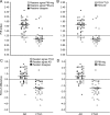
Results: PiB visual reads had a higher sensitivity for AD (89.5% average between raters) than FDG visual reads (77.5%) with similar specificity (PiB 83%, FDG 84%). When scans were classified quantitatively, PiB had higher sensitivity (89% vs 73%) while FDG had higher specificity (83% vs 98%). On receiver operating characteristic analysis, areas under the curve for PiB (0.888) and FDG (0.910) were similar. Interrater agreement was higher for PiB (κ = 0.96) than FDG (κ = 0.72), as was agreement between visual and quantitative classification (PiB κ = 0.88-0.92; FDG κ = 0.64-0.68). In patients with known histopathology, overall classification accuracy (2 visual and 1 quantitative classification per patient) was 97% for PiB (n = 12 patients) and 87% for FDG (n = 10).
Conclusions: PiB and FDG showed similar accuracy in discriminating AD and FTLD. PiB was more sensitive when interpreted qualitatively or quantitatively. FDG was more specific, but only when scans were classified quantitatively. PiB slightly outperformed FDG in patients with known histopathology.
Figures


Comment in
-
A tale of two tracers: the age of wisdom for dementia diagnosis?Neurology. 2011 Dec 6;77(23):2008-9. doi: 10.1212/WNL.0b013e31823b9c9f. Epub 2011 Nov 30. Neurology. 2011. PMID: 22131545 No abstract available.
References
-
- Mendez MF, Shapira JS, McMurtray A, Licht E. Preliminary findings: behavioral worsening on donepezil in patients with frontotemporal dementia. Am J Geriatr Psychiatry 2007;15:84–87 - PubMed
-
- Roberson ED, Hesse JH, Rose KD, et al. Frontotemporal dementia progresses to death faster than Alzheimer disease. Neurology 2005;65:719–725 - PubMed
-
- Galton CJ, Patterson K, Xuereb JH, Hodges JR. Atypical and typical presentations of Alzheimer's disease: a clinical, neuropsychological, neuroimaging and pathological study of 13 cases. Brain 2000;123:484–498 - PubMed
-
- Graham A, Davies R, Xuereb J, et al. Pathologically proven frontotemporal dementia presenting with severe amnesia. Brain 2005;128:597–605 - PubMed
MeSH terms
Substances
Grants and funding
LinkOut - more resources
Full Text Sources
Medical
