

Photoreceptor disruption related to persistent submacular fluid after successful scleral buckle surgery
- PMID: 22131774
- PMCID: PMC3223704
- DOI: 10.3341/kjo.2011.25.6.380
Photoreceptor disruption related to persistent submacular fluid after successful scleral buckle surgery
Abstract
Purpose: To investigate serial changes in photoreceptor status and associated visual outcome in patients with persistent submacular fluid after successful scleral buckle surgery for macula-off rhegmatogenous retinal detachment.
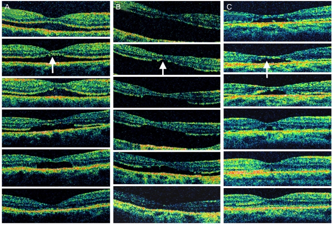
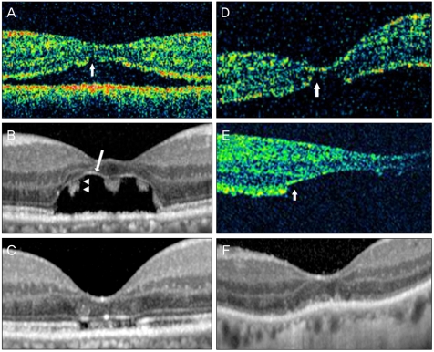
Methods: This was a prospective observational case series including 76 consecutive patients who underwent successful scleral buckle surgery for macula-off rhegmatogenous retinal detachment with symptom duration ≤90 days at a single tertiary hospital. Optical coherence tomography (OCT) and visual acuity examination were performed at one month and three months postoperatively and at three-month intervals until the submacular fluid disappeared. Main outcome measures were postoperative photoreceptor status on OCT and visual acuity.
Results: Forty-two patients (55.3%) showed persistent submacular fluid at postoperative one month. Of 42 patients with persistent submacular fluid, three (7.1%) showed photoreceptor disruption on OCT. None of the 34 patients without persistent submacular fluid showed photoreceptor disruption. Two patients (4.8%) had progressive photoreceptor disruption, and one patient (2.4%) had early photoreceptor disruption. All three patients showed photoreceptor reappearance and limited visual restoration after absorption of submacular fluid. Final visual acuities were significantly worse in these three patients (20 / 1000, 20 / 133, and 20 / 133) compared to those of the other patients (mean, 20 / 30) with persistent submacular fluid and intact photoreceptors.
Conclusions: Even after successful scleral buckle surgery for rhegmatogenous retinal detachment, photoreceptor disruption can occur related to persistent submacular fluid and may be a cause of poor visual outcome.
Keywords: Optical coherence tomography; Photoreceptor cells; Retinal detachment; Sclera buckling; Subretinal fluid.
Conflict of interest statement
No potential conflict of interest relevant to this article was reported.
Figures


Similar articles
-
Influence of persistent submacular fluid on visual outcome after successful scleral buckle surgery for macula-off retinal detachment.Am J Ophthalmol. 2008 May;145(5):915-22. doi: 10.1016/j.ajo.2008.01.005. Epub 2008 Mar 10. Am J Ophthalmol. 2008. PMID: 18329625
-
Optical coherence tomography analysis of the macula after scleral buckle surgery for retinal detachment.Ophthalmology. 2007 Jan;114(1):108-12. doi: 10.1016/j.ophtha.2006.07.022. Epub 2006 Nov 7. Ophthalmology. 2007. PMID: 17095091
-
[Influencing factors of visual prognosis in patients with persistent submacular fluid after successful scleral buckle surgery for macula-off retinal detachment].Zhonghua Yan Ke Za Zhi. 2023 Nov 11;59(11):899-905. doi: 10.3760/cma.j.cn112142-20230809-00030. Zhonghua Yan Ke Za Zhi. 2023. PMID: 37936358 Chinese.
-
Persistent subretinal fluid after surgery for rhegmatogenous retinal detachment: hypothesis and review.Graefes Arch Clin Exp Ophthalmol. 2012 Jun;250(6):795-802. doi: 10.1007/s00417-011-1870-y. Epub 2012 Jan 11. Graefes Arch Clin Exp Ophthalmol. 2012. PMID: 22234351 Review.
-
Persistent Subretinal Fluid Following Successful Rhegmatogenous Retinal Detachment Surgery.Semin Ophthalmol. 2022 Aug;37(6):724-729. doi: 10.1080/08820538.2022.2085516. Epub 2022 Jun 6. Semin Ophthalmol. 2022. PMID: 35666630 Review.
Cited by
-
Spontaneous resolution of subretinal fluid postvitreoretinal surgery for tractional retinal detachment in proliferative diabetic retinopathy.BMJ Case Rep. 2022 Aug 25;15(8):e249745. doi: 10.1136/bcr-2022-249745. BMJ Case Rep. 2022. PMID: 36007977 Free PMC article.
-
Mineralocorticoid receptor antagonists as a potential treatment option in persistent subretinal fluid following the repair of a rhegmatogenous retinal detachment.Am J Ophthalmol Case Rep. 2022 Dec 31;29:101789. doi: 10.1016/j.ajoc.2022.101789. eCollection 2023 Mar. Am J Ophthalmol Case Rep. 2022. PMID: 36718434 Free PMC article.
-
Micropulse Laser for Persistent Sub-Retinal Fluid in a Patient Previously Treated for Rhegmatogenous Retinal Detachment.Med Hypothesis Discov Innov Ophthalmol. 2018 Winter;7(4):190-194. Med Hypothesis Discov Innov Ophthalmol. 2018. PMID: 30505871 Free PMC article.
-
Multifactor analysis of delayed absorption of subretinal fluid after scleral buckling surgery.BMC Ophthalmol. 2021 Feb 15;21(1):86. doi: 10.1186/s12886-021-01853-2. BMC Ophthalmol. 2021. PMID: 33588767 Free PMC article.
References
-
- Wolfensberger TJ, Gonvers M. Optical coherence tomography in the evaluation of incomplete visual acuity recovery after macula-off retinal detachments. Graefes Arch Clin Exp Ophthalmol. 2002;240:85–89. - PubMed
-
- Seo JH, Woo SJ, Park KH, et al. Influence of persistent submacular fluid on visual outcome after successful scleral buckle surgery for macula-off retinal detachment. Am J Ophthalmol. 2008;145:915–922. - PubMed
-
- Hagimura N, Iida T, Suto K, Kishi S. Persistent foveal retinal detachment after successful rhegmatogenous retinal detachment surgery. Am J Ophthalmol. 2002;133:516–520. - PubMed
-
- Benson SE, Schlottmann PG, Bunce C, et al. Optical coherence tomography analysis of the macula after scleral buckle surgery for retinal detachment. Ophthalmology. 2007;114:108–112. - PubMed
-
- Ross WH. Visual recovery after macula-off retinal detachment. Eye (Lond) 2002;16:440–446. - PubMed
Publication types
MeSH terms
LinkOut - more resources
Full Text Sources
Medical
