

Keeping the balance between proliferation and differentiation: the primary cilium
- PMID: 22131874
- PMCID: PMC3131736
- DOI: 10.2174/138920211795860134
Keeping the balance between proliferation and differentiation: the primary cilium
Abstract
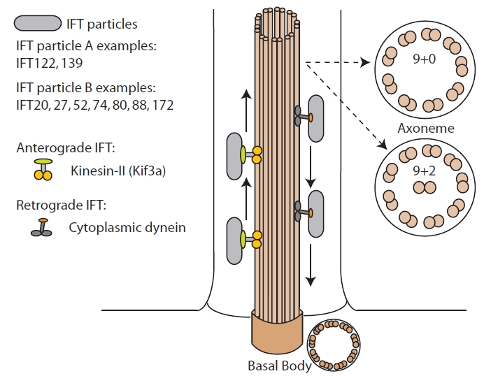
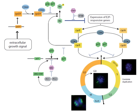
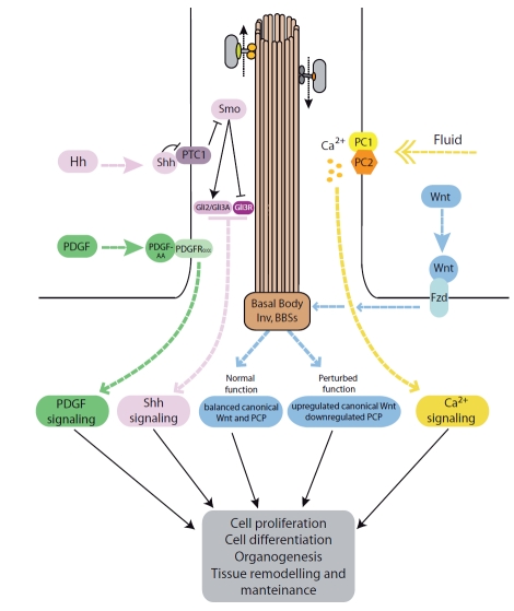
Primary cilia are post-mitotic cellular organelles that are present in the vast majority of cell types in the human body. An extensive body of data gathered in recent years is demonstrating a crucial role for this organelle in a number of cellular processes that include mechano and chemo-sensation as well as the transduction of signaling cascades critical for the development and maintenance of different tissues and organs. Consequently, cilia are currently viewed as cellular antennae playing a critical role at the interphase between cells and their environment, integrating a range of stimuli to modulate cell fate decisions including cell proliferation, migration and differentiation. Importantly, this regulatory role is not just a consequence of their participation in signal transduction but is also the outcome of both the tight synchronization/regulation of ciliogenesis with the cell cycle and the role of individual ciliary proteins in cilia-dependent and independent processes. Here we review the role of primary cilia in the regulation of cell proliferation and differentiation and illustrate how this knowledge has provided insight to understand the phenotypic consequences of ciliary dysfunction.
Keywords: Cilia; cancer.; cell cycle; ciliopathies.
Figures
Similar articles
-
Primary Cilia, Ciliogenesis and the Actin Cytoskeleton: A Little Less Resorption, A Little More Actin Please.Front Cell Dev Biol. 2020 Dec 17;8:622822. doi: 10.3389/fcell.2020.622822. eCollection 2020. Front Cell Dev Biol. 2020. PMID: 33392209 Free PMC article. Review.
-
Ciliary biology: understanding the cellular and genetic basis of human ciliopathies.Am J Med Genet C Semin Med Genet. 2009 Nov 15;151C(4):263-80. doi: 10.1002/ajmg.c.30227. Am J Med Genet C Semin Med Genet. 2009. PMID: 19876935 Review.
-
[Current situation of researches on a sensor organelle, primary cilium, to understand the pathogenesis of ciliopathy].Nihon Yakurigaku Zasshi. 2019;153(3):117-123. doi: 10.1254/fpj.153.117. Nihon Yakurigaku Zasshi. 2019. PMID: 30867380 Japanese.
-
Functional aspects of primary cilium in signaling, assembly and microenvironment in cancer.J Cell Physiol. 2021 May;236(5):3207-3219. doi: 10.1002/jcp.30117. Epub 2020 Oct 26. J Cell Physiol. 2021. PMID: 33107052 Free PMC article. Review.
-
Genes and molecular pathways underpinning ciliopathies.Nat Rev Mol Cell Biol. 2017 Sep;18(9):533-547. doi: 10.1038/nrm.2017.60. Epub 2017 Jul 12. Nat Rev Mol Cell Biol. 2017. PMID: 28698599 Free PMC article. Review.
Cited by
-
Primary cilia and kidney injury: current research status and future perspectives.Am J Physiol Renal Physiol. 2013 Oct 15;305(8):F1085-98. doi: 10.1152/ajprenal.00399.2013. Epub 2013 Jul 31. Am J Physiol Renal Physiol. 2013. PMID: 23904226 Free PMC article. Review.
-
Primary cilia suppress the fibrotic activity of atrial fibroblasts from patients with atrial fibrillation in vitro.Sci Rep. 2024 May 30;14(1):12470. doi: 10.1038/s41598-024-60298-x. Sci Rep. 2024. PMID: 38816374 Free PMC article.
-
A mouse model of Bardet-Biedl Syndrome has impaired fear memory, which is rescued by lithium treatment.PLoS Genet. 2021 Apr 22;17(4):e1009484. doi: 10.1371/journal.pgen.1009484. eCollection 2021 Apr. PLoS Genet. 2021. PMID: 33886537 Free PMC article.
-
Novel insights into the genetic landscape of congenital heart disease with systems genetics.Prog Pediatr Cardiol. 2019 Sep;54:101128. doi: 10.1016/j.ppedcard.2019.101128. Epub 2019 Jul 10. Prog Pediatr Cardiol. 2019. PMID: 34404969 Free PMC article.
-
IFT80 is essential for chondrocyte differentiation by regulating Hedgehog and Wnt signaling pathways.Exp Cell Res. 2013 Mar 10;319(5):623-32. doi: 10.1016/j.yexcr.2012.12.028. Epub 2013 Jan 16. Exp Cell Res. 2013. PMID: 23333501 Free PMC article.
References
-
- Rosenbaum JL, Witman GB. Intraflagellar transport. Nat. Rev. Mol. Cell Biol. 2002;3:813–825. - PubMed
-
- Cardenas-Rodriguez M, Badano JL. Ciliary biology: understanding the cellular and genetic basis of human ciliopathies. Am. J. Med. Genet. C Semin. Med. Genet. 2009;151C:263–280. - PubMed
-
- Pedersen LB, Rosenbaum JL. Intraflagellar transport (IFT) role in ciliary assembly, resorption and signalling. Curr. Top. Dev. Biol. 2008;85:23–61. - PubMed
-
- Badano JL, Mitsuma N, Beales PL, Katsanis N. The ciliopathies: An emergin class of human genetic disorders. Annu. Rev. Genomics Hum. Genet. 2006;7:125–148. - PubMed
LinkOut - more resources
Full Text Sources