. 2011 Nov;13(11):1069-80.
doi: 10.1593/neo.111032.

DNA-PK mediates AKT activation and apoptosis inhibition in clinically acquired platinum resistance

Affiliations

DNA-PK mediates AKT activation and apoptosis inhibition in clinically acquired platinum resistance

Euan A Stronach et al. Neoplasia. 2011 Nov.

Abstract

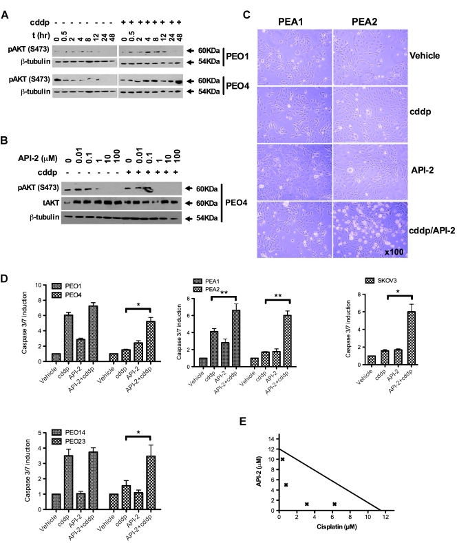
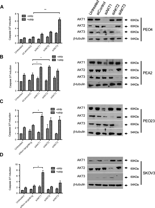
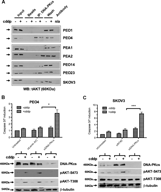
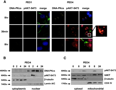
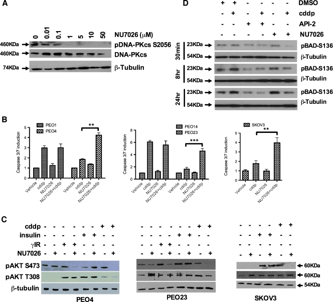
Clinical resistance to chemotherapy is a frequent event in cancer treatment and is closely linked to poor outcome. High-grade serous (HGS) ovarian cancer is characterized by p53 mutation and high levels of genomic instability. Treatment includes platinum-based chemotherapy and initial response rates are high; however, resistance is frequently acquired, at which point treatment options are largely palliative. Recent data indicate that platinum-resistant clones exist within the sensitive primary tumor at presentation, implying resistant cell selection after treatment with platinum chemotherapy. The AKT pathway is central to cell survival and has been implicated in platinum resistance. Here, we show that platinum exposure induces an AKT-dependent, prosurvival, DNA damage response in clinically platinum-resistant but not platinum-sensitive cells. AKT relocates to the nucleus of resistant cells where it is phosphorylated specifically on S473 by DNA-dependent protein kinase (DNA-PK), and this activation inhibits cisplatin-mediated apoptosis. Inhibition of DNA-PK or AKT, but not mTORC2, restores platinum sensitivity in a panel of clinically resistant HGS ovarian cancer cell lines: we also demonstrate these effects in other tumor types. Resensitization is associated with prevention of AKT-mediated BAD phosphorylation. Strikingly, in patient-matched sensitive cells, we do not see enhanced apoptosis on combining cisplatin with AKT or DNA-PK inhibition. Insulin-mediated activation of AKT is unaffected by DNA-PK inhibitor treatment, suggesting that this effect is restricted to DNA damage-mediated activation of AKT and that, clinically, DNA-PK inhibition might prevent platinum-induced AKT activation without interfering with normal glucose homeostasis, an unwanted toxicity of direct AKT inhibitors.


Figures

Figure 1
Figure 1
AKT inhibition reverses resistance to platinum treatment in ovarian cancer cells. Western blot indicates differential AKT-S473 response to platinum treatment between platinum-sensitive and -resistant cells from the same patient (A) and shows inhibition of pAKT by AKT inhibitor API-2 (B). Visual appearance of platinum-sensitive (PEA1) and matched resistant (PEA2) cells illustrates enhancement of cytotoxicity in resistant cells 8 hours after combination treatment with 20 µM API-2 and equitoxic cisplatin (5 µM PEA1; 25 µM PEA2) (C). Caspase 3/7 assays 24 hours after treatment with cisplatin and/or API-2 reveal enhanced apoptosis when combining API-2 with cisplatin compared to platinum alone in platinum-resistant cell lines: PEO4 (P = .019), PEA2 (P = .003), PEO23 (P = .042), and SKOV3 (P = .02) (paired t tests). Conversely, platinum-induced apoptosis is more modestly enhanced in the matched platinum-sensitive cells lines PEO1, PEA1, and PEO14, with only PEA1 achieving statistical significance (P = .01) (D). Isobologram analysis of acquired platinum-resistant PEO4 cells supports the synergistic interaction between platinum and API-2 (E). n ≥ 3. *P < .05. **P < .01.
Figure 2
Figure 2
The functional effect of AKT in platinum resistance is not determined by a single common AKT isoform. After knockdown of individual AKT isoforms 1, 2, and 3, cells were treated with cisplatin, and caspase 3/7 activation was assayed for platinum-resistant cell lines PEO4 (A), PEA2 (B), PEO23 (C), and SKOV3 (D). Isoform-specific knockdowns are indicated by Western blot (right panels). Statistically significant differences, compared with control transfections, are indicated: *P < .05. **P < .01.
Figure 3
Figure 3
AKT is not activated by mTOR-Rictor in response to platinum, and its inhibition does not enhance platinum-mediated apoptosis. Caspase 3/7 activation was measured 24 hours after cisplatin treatment in control and Rictor siRNA-transfected cells, which indicated no enhancement of apoptosis or effect on pAKT-S473 (A). Treatment of sensitive (PEO1) and resistant (PEO4) cells with the mTOR inhibitor rapamycin (200 nM) and/or cisplatin (25 µM) indicated no enhancement of platinum sensitivity on combination with rapamycin and no effect on AKT phosphorylation (B). No interaction between Rictor and pAKT was detected by IP in the presence or absence of 25 µM cisplatin (2-hour exposure) in platinum-resistant PEO4 cells (C).
Figure 4
Figure 4
DNA-PKcs phosphorylates AKT in response to cisplatin treatment and mediates platinum resistance. IP of DNA-PKcs in a panel of platinum-sensitive and -resistant cells indicates interaction with AKT in resistant cells, in the presence and absence of 25 µM cisplatin (2-hour exposure), which is not seen in sensitive PEO1 and PEA1 cells and is only detectable on platinum treatment in sensitive PEO14 cells (A). Knockdown of DNA-PKcs in platinum-resistant PEO4 (B) and SKOV3 (C) cells increases the apoptotic induction on platinum treatment compared with control transfections (P = .01, PEO4: P = .0003, SKOV3). Western blots indicate specific loss of cisplatin-induced phosphorylation of AKT-S473 but not T308. n = 3. *P < .05. ***P < .001.
Figure 5
Figure 5
After platinum treatment, AKT is translocated to the nucleus of cisplatin-resistant but not matched cisplatin-sensitive cells. Immunofluorescent microscopy of platinum-sensitive (PEO1) and -resistant (PEO4) cells after treatment with 25 µM cisplatin reveals induction and nuclear accumulation of pAKT-S473 in PEO4 but not PEO1. The enlarged box indicates nuclear colocalization of pAKT and DNA-PKcs in PEO4 cells 30 minutes after platinum treatment. After an 8-hour exposure to cisplatin, pAKT appears redistributed to the cytoplasmic compartment in PEO4. Counterstaining of nuclei is indicated in blue in the merged images (secondary antibody-only controls are shown in Figure W4; A). Western blot of fractionated PEO4 cells confirms early nuclear location of pAKT after 25 µM cisplatin with delayed cytoplasmic accumulation. Purity of fractions is indicated by β-tubulin (cytoplasmic) and Lamin A/C (nuclear) markers (B). Mitochondrial fractionation indicates that punctate staining seen 8 hours after cisplatin treatment in A corresponds with a mitochondrial relocalization of pAKT. Purity of fractions is indicated by β-tubulin (cytoplasmic) and COX IV (mitochondrial) markers (C).
Figure 6
Figure 6
DNA-PK inhibition enhances cisplatin-induced apoptosis in platinum-resistant but not platinum-sensitive cells through inhibition of AKT-S473 phosphorylation. Inhibition of DNA-PKcs phosphorylation at serine 2056 was observed from 1 to 50 µM DNA-PK inhibitor NU7026, consistent with DNA-PKcs autophosphorylation at this residue (A). Forty-eight hours of cisplatin treatment of intrapatient paired platinum-sensitive (PEO1 and PEO14; 5 µM cisplatin) and -resistant (PEO4 and PEO23; 25 µM cisplatin) after serum starvation indicates clear and significant resensitization to platinum-mediated apoptosis after 10 µM NU7026 treatment in resistant cells (P = .0017, PEO4; P = .0004, PEO23) but no enhancement of apoptosis in sensitive cells (P = n.s.). The unmatched platinum-resistant SKOV3 cell line behaves similarly to the other resistant lines (P = .001) (B). Western blot indicates a consistent inhibition of pAKT-S473 in platinum- or IR-treated cells when cotreated with NU7026 for 24 hours. Conversely, AKT T308 phosphorylation is unaffected by NU7026 treatment. DNA-PK inhibition failed to prevent insulin-mediated AKT-S473 phosphorylation (C). Western blot indicates inhibition of pBAD-S136 at 30 minutes, 8 hours, and 24 hours after combined treatment with cisplatin (25 µM) and the AKT inhibitor (API-2; 10 µM) or DNA-PK inhibitor (NU7026; 10 µM). (D) **P < .01. ***P < .001.

Similar articles

Cited by

References

Supplementary References

    1. Stronach EA, Alfraidi A, Rama N, Datler C, Studd JB, Agarwal R, Guney TG, Gourley C, Hennessy BT, Mills GB, et al. HDAC4-regulated STAT1 activation mediates platinum resistance in ovarian cancer. Cancer Res. 2011;71(13):4412–4422. - PMC - PubMed
    1. Goh LB, Spears KJ, Yao D, Ayrton A, Morgan P, Roland Wolf C, Friedberg T. Endogenous drug transporters in in vitro and in vivo models for the prediction of drug disposition in man. Biochem Pharmacol. 2002;64(11):1569–1578. - PubMed
    1. Shahbazi M, Fryer AA, Pravica V, Brogan IJ, Ramsay HM, Hutchinson IV, Harden PN. Vascular endothelial growth factor gene polymorphisms are associated with acute renal allograft rejection. J Am Soc Nephrol. 2002;13(1):260–264. - PubMed

References

    1. Vivanco I, Sawyers CL. The phosphatidylinositol 3-kinase AKT pathway in human cancer. Nat Rev Cancer. 2002;2:489–501. - PubMed
    1. Fraser M, Leung BM, Yan X, Dan HC, Cheng JQ, Tsang BK. p53 is a determinant of X-linked inhibitor of apoptosis protein/Akt-mediated chemoresistance in human ovarian cancer cells. Cancer Res. 2003;63:7081–7088. - PubMed
    1. Yang X, Fraser M, Moll UM, Basak A, Tsang BK. Akt-mediated cisplatin resistance in ovarian cancer: modulation of p53 action on caspase-dependent mitochondrial death pathway. Cancer Res. 2006;66:3126–3136. - PubMed
    1. Molina JR, Hayashi Y, Stephens C, Georgescu MM. Invasive glioblastoma cells acquire stemness and increased Akt activation. Neoplasia. 2010;12:453–463. - PMC - PubMed
    1. Blando JM, Carbajal S, Abel E, Beltran L, Conti C, Fischer S, DiGiovanni J. Cooperation between Stat3 and Akt signaling leads to prostate tumor development in transgenic mice. Neoplasia. 2011;13:254–265. - PMC - PubMed

Publication types

MeSH terms

LinkOut - more resources