. 2011 Nov;9(11):e1001202.
doi: 10.1371/journal.pbio.1001202. Epub 2011 Nov 22.

Coordinated regulation of niche and stem cell precursors by hormonal signaling

Affiliations

Coordinated regulation of niche and stem cell precursors by hormonal signaling

Dana Gancz et al. PLoS Biol. 2011 Nov.

Abstract

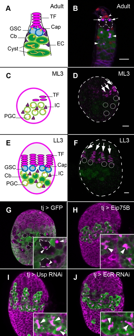
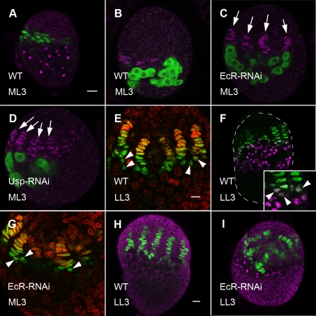
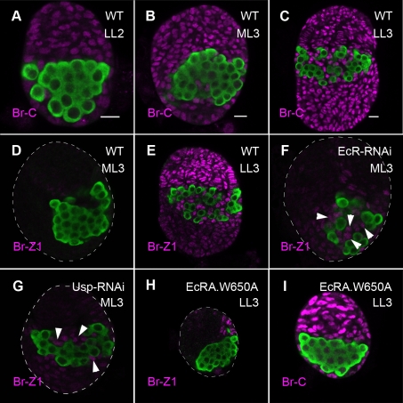
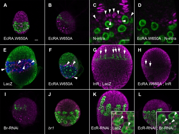
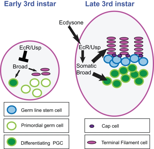
Stem cells and their niches constitute units that act cooperatively to achieve adult body homeostasis. How such units form and whether stem cell and niche precursors might be coordinated already during organogenesis are unknown. In fruit flies, primordial germ cells (PGCs), the precursors of germ line stem cells (GSCs), and somatic niche precursors develop within the larval ovary. Together they form the 16-20 GSC units of the adult ovary. We show that ecdysone receptors are required to coordinate the development of niche and GSC precursors. At early third instar, ecdysone receptors repress precocious differentiation of both niches and PGCs. Early repression is required for correct morphogenesis of the ovary and for protecting future GSCs from differentiation. At mid-third instar, ecdysone signaling is required for niche formation. Finally, and concurrent with the initiation of wandering behavior, ecdysone signaling initiates PGC differentiation by allowing the expression of the differentiation gene bag of marbles in PGCs that are not protected by the newly formed niches. All the ovarian functions of ecdysone receptors are mediated through early repression, and late activation, of the ecdysone target gene broad. These results show that, similar to mammals, a brain-gland-gonad axis controls the initiation of oogenesis in insects. They further exemplify how a physiological cue coordinates the formation of a stem cell unit within an organ: it is required for niche establishment and to ensure that precursor cells to adult stem cells remain undifferentiated until the niches can accommodate them. Similar principles might govern the formation of additional stem cell units during organogenesis.


Conflict of interest statement

The authors have declared that no competing interests exist.

Figures

Figure 1
Figure 1. Ecdysone receptors repress precocious PGC differentiation.
(A) An illustration of an adult germarium. Niche cells (Terminal Filament, TF, and Cap cells) are at the anterior (magenta). Attached to cap cells are the Germ Line Stem Cells (GSCs, blue). GSC progeny (Cystoblast, Cb; and cysts, green) are posterior to GSCs. GSCs and Cbs carry a round fusome (yellow), while germ line cysts carry a branched fusome. Germ cells are also contacted by somatic Escort Cells (ECs, purple). (B) Adult germarium. TF and cap cells (barbed arrowheads) are marked by hedgehog-lacZ (anti β-Galactosidase, red). GSCs (outlined) are attached to cap cells. Fusomes within GSCs are labeled by monoclonal antibody 1B1 (magenta) and are round (arrows). Posterior to GSCs, differentiating germ cells are expressing bamP-GFP (green). Fusomes within germ line cysts are extended or branched (arrowheads). (C) An illustration of a larval ovary at mid-3rd instar (ML3). TF cells (magenta) are only beginning to form. Primordial germ cells (PGCs, green) are more posterior. Intermingled Cells (ICs, purple) are associated with PGCs. (D) An ML3 ovary. Terminal filament cells are labeled by anti-Engrailed (En, magenta). Few En expressing cells are present in very short filaments (arrows). Differentiating PGCs are labeled by bamP-GFP (anti-GFP, green). PGCs have not yet differentiated, and cannot be recognized by anti-GFP; their location is indicated by white circles. (E) An illustration of a late 3rd (LL3) ovary. Somatic niches (TF, Cap cells, magenta) are marked. GSCs (blue) are established close to TF and Cap cells. Away from niches, PGCs initiate differentiation and some have turned to cystoblasts (dark green). (F) An LL3 ovary. TF stacks and cap cells, marked by anti-En (magenta, arrows), are formed throughout the anterior of the ovary. Many PGCs that are not close to the niches are expressing bamP-GFP (anti-GFP, green). PGCs that become GSCs are close to niches. They do not express bamP-GFP and their location is indicated by white circles. (G–J) LL3 ovaries, all labeled by 1B1 antibody to outline somatic cells and fusomes within PGCs (magenta). (G) The somatic driver tj-Gal4 drives GFP expression (anti-GFP, green) in somatic cells, but not in PGCs (some PGCs are outlined, inset). PGCs in wild-type LL3 ovaries carry round fusomes (inset, arrowheads). (H–J) PGCs are labeled by anti-Vasa (green). Somatic expression of Eip75B (H) or RNAi construct against usp (I) or RNAi construct against EcR (J) results in formation of multiple cysts harboring branched fusomes (arrowheads, insets). Bars in panel (B), (D), and (F) (for F–J) are 10 µm. Anterior is up.
Figure 2
Figure 2. Ecdysone receptors repress precocious niche formation.
(A, B) Terminal filaments of wild-type ML3 ovaries are labeled either by hh-lacZ (A, green) or anti-En (B, magenta). Few TF cells which are unorganized, or organized into short filaments, can be seen. Germ cells are marked by round fusomes (A, 1B1 antibody, magenta) or by anti-Vasa (B, green). (C, D) In EcR-RNAi (C) or usp-RNAi (D) ML3 ovaries, more TF cells and more organized filaments can be seen (anti-En, magenta, arrows). Germ cells are labeled by anti-Vasa (green). (E–I) hh-lacZ (green) marks all TF and cap cells. (E) In WT LL3 ovaries TF cells are distinguished by hh-lacZ staining and oval-shaped nuclei. LaminC (red) is only apparent in older TF cells, at the anterior of each TF stack. Cap cells (arrowheads) are at the posterior base of TF, have rounder nuclei, and do not yet stain with anti-laminC. (F) Anti-Tj (magenta) labels ICs. Cap cells that form at the base of TFs are co-stained with hh-LacZ and anti-Tj (inset, arrowheads). (G) EcR-RNAi ML3 ovaries. Unlike wild-type, Cap cells appear already at ML3 (arrowheads). (H, I) Somatic cells and fusomes are labeled by 1B1 (magenta). (H) TFs are regularly spaced in the anterior half of the wild-type ovary. (I) In EcR-RNAi LL3 ovaries, TFs are mis-positioned, with fewer cells between stacks. Bars in (A), (for A–D), in E (for E, G), and in H (for F, H, I) are 10 µm.
Figure 3
Figure 3. Somatic ecdysone receptors are early repressors and late activators of Broad-Z1.
In all panels, germ cells are labeled with anti-Vasa (green). Panels (A–C) were taken with the same confocal settings. Antibodies against the common region of Broad (Broad-C, magenta) stain somatic cell nuclei of LL2 (A), ML3 (B), and LL3 (C) ovaries. Staining levels become stronger with time. Panels (D–G) were taken with the same confocal settings. Broad-Z1 (magenta) is very weakly expressed in wild type ML3 ovaries (D), but is strongly expressed at LL3 (E). In contrast to wild type, Br-Z1 is expressed in ICs (arrowheads) of EcR-RNAi (F) and usp-RNAi (G) ML3 ovaries. (H) Broad-Z1 (magenta) is not expressed in most somatic cells of EcRA.W650A ovaries. (I) In contrast, anti-Br-C does label somatic cells of EcRA.W650A ovaries. Bars in (A), in (B) (for B, D, F, G), and in (C) (C, E, H, I) are 10 µm.
Figure 4
Figure 4. Gonad development requires ecdysone signaling and Broad expression.
In all panels except (C, D), 1B1 antibody labels fusomes and outlines somatic cells. (A) Anti-Vasa (green) labels PGCs. EcRA.W650A LL3 ovaries are much smaller than wild type ovaries (compare Figure 4A to Figure 1G). (B) EcRA.W650A ovaries contain very few TF cells (hh-LacZ, green), which are not organized into stacks (compare Figure 4B to Figure 2H). (C, D) TF and cap cells are labeled by anti-En (magenta). PGCs are labeled by anti-Vasa (green). (C) Expression of N-intra greatly increases the number of En-labeled cap cells at the base of TFs (arrowheads). (D) Cap cells are not observed in EcRA.W650A ovaries. (E, F) ICs are labeled by anti-Tj (magenta) and are located next to PGCs (anti-Vasa, Blue). 1B1 labeling is green. (E) In wild-type, no Tj labeling is observed at the anterior at LL3. ICs intermingle with PGCs. (F) In EcRA.W650A ovaries, ICs lie outside of the PGC region. As in wild type, no Tj staining is observed at the anterior. (G–I) TFs are labeled by hh-lacZ (green). (G) Ovaries over-expressing InR are larger and contain fully formed TF and cap cells (arrows). (H) Over-expression of InR in EcRA.W650A rescues gonadal size. However, very few TF cells are specified (arrows), which are not organized into stacks. (I) Fewer, shorter TFs are present in br-RNAi ovaries. (J) br1 LL3 ovaries are small with less developed TFs. (K, L) Precocious cysts and TFs in EcR-RNAi ovaries (K, inset, arrowheads) are not observed when ovaries also lack broad (I, inset, arrowheads). Bars in (A) (for A, B, E–L) and in C (for C, D) are 10 µm.
Figure 5
Figure 5. Broad over-expression results in precocious niche and PGC differentiation.
(A–C) TF cells are marked by anti-Engrailed (magenta). Differentiating PGCs are marked by bamP-GFP (anti-GFP, green). (A) In wild type ML3 ovaries, TF formation initiates, but PGCs are not yet differentiating. No GFP labeling is evident. (B, C) In Br-Z1 (B) and Br-Z4 (C) over-expressing ML3 ovaries, substantial PGC differentiation is observed. More TF cells are specified in Br-Z1, but not in Br-Z4 ovaries. (D–F) PGCs are labeled green (anti-Vasa). (D) In a wild-type adult germarium, 8- and 16-cell cysts are labeled with anti-Orb (magenta). Orb localizes to the selected oocyte (arrowheads). (E) Wild-type PGCs do not express Orb. (F) Cysts in Br-Z1 over-expressing ovaries can express Orb, which is sometimes localized to an oocyte (arrowheads). Bars in (A) (for A–C), in (D), and in (E) (for E, F) are 10 µm.
Figure 6
Figure 6. Ecdysone signaling is required for bam up-regulation in differentiating PGCs.
All panels depict LL3 ovaries. 1B1 (magenta) outlines somatic cells and fusomes. (A) In wild type LL3 ovaries, only PGCs closest to the niche retain pMad labeling (anti-pMad, green). (A′) is a grayscale image of the green channel. PGC region is outlined. pMAD positive PGC nuclei are marked by arrowheads. (B) PGCs that are located away from the niche and do not carry pMAD in their nuclei up-regulate bam (bamP-GFP, green). PGCs located in niches do not express bamP-GFP (outlined). (C) In EcRA.W650A ovaries, some PGCs, which are close to anterior or posterior somatic cells, retain pMAD labeling. Most PGCs do not retain pMad staining. (C′) is a grayscale image of the green channel. PGC region is outlined. pMAD positive nuclei are marked by arrowheads. (D) No corresponding elevation of bamP-GFP can be observed in EcRA.W650A ovaries. Bar in (A) for all panels is 10 µm.
Figure 7
Figure 7. Distinct ecdysone-mediated events are required for niche formation and for PGC differentiation.
(A–D) Niche cells are labeled with Anti-En (magenta). (A, B) PGCs containing pMAD (anti-pMAD, green, arrowheads) can be seen in both control and EcRA.W650A ovaries, close to newly formed cap cells (arrows). (C, D) Differentiating PGCs express bamP-GFP (anti-GFP, green). (C) In wild-type LL3 ovaries, PGCs that are not associated with niches differentiate and express bamP-GFP normally. (D) When ecdysone signaling is blocked by expression of EcRA.W650A after niches form, but prior to PGC differentiation, PGCs fail to differentiate despite normal TF formation. No GFP expression in PGCs is observed. Faint, non-specific, GFP can be observed in a few TF cells in both control and experimental ovaries. (E) Br-Z1 (anti Br-Z1, magenta) is expressed in formed niches of EcRA.W650A temperature-shifted ovaries (arrows), but is not expressed in ICs. Bar in (A) (for A–E) is 10 µm. (F) 0–4 h prior to wandering behavior (in food), most ovaries do not carry differentiating PGCs (light bars), while 0–4 h following the initiation of wandering, most ovaries carry differentiating PGCs (black bars).
Figure 8
Figure 8. Coordination of niche formation with GSC establishment by ecdysone.
At early third instar, ecdysone receptors repress niche formation and PGC differentiation; this allows the gonad time to grow and generate sufficient precursor cells of both cell populations that will eventually form 16–20 stem cell units. Repression of the target gene broad is a key component of this repression. At mid-third instar and later, the hormone ecdysone activates Broad-Z1 expression in the somatic cells of the ovary through EcR and Usp. This leads first to formation of niches and, later, concomitant with wandering behavior, to PGC differentiation, by an unknown mechanism. Only PGCs that are located next to niches are protected from differentiation and become the adult GSCs.

Similar articles

Cited by

References

    1. Fuller M. T, Spradling A. C. Male and female Drosophila germline stem cells: two versions of immortality. Science. 2007;316:402–404. - PubMed
    1. Kirilly D, Xie T. The Drosophila ovary: an active stem cell community. Cell Res. 2007;17:15–25. - PubMed
    1. Xie T, Spradling A. C. decapentaplegic is essential for the maintenance and division of germline stem cells in the Drosophila ovary. Cell. 1998;94:251–260. - PubMed
    1. Xie T, Spradling A. C. A niche maintaining germ line stem cells in the Drosophila ovary. Science. 2000;290:328–330. - PubMed
    1. Chen D, McKearin D. Dpp signaling silences bam transcription directly to establish asymmetric divisions of germline stem cells. Curr Biol. 2003;13:1786–1791. - PubMed

Publication types

MeSH terms