

Bifid thumb type IV in children: transferring an epiphyseal segment of the proximal phalanx with insertion of the abductor pollicis brevis tendon
- PMID: 22132030
- PMCID: PMC2981707
- DOI: 10.1007/s11832-010-0296-0
Bifid thumb type IV in children: transferring an epiphyseal segment of the proximal phalanx with insertion of the abductor pollicis brevis tendon
Abstract
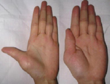
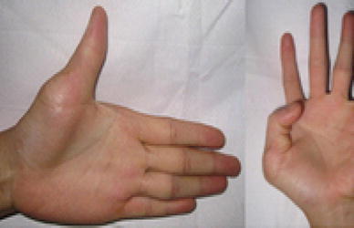
Objective: To evaluate the clinical and functional results of the surgical treatment of bifid thumb type IV in children.
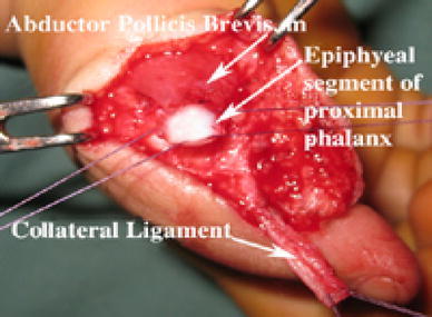
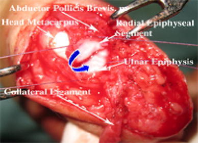
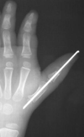
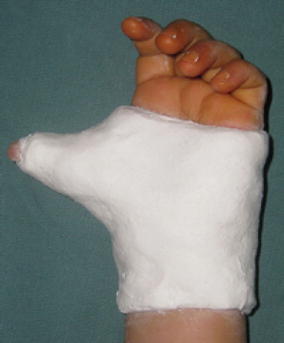
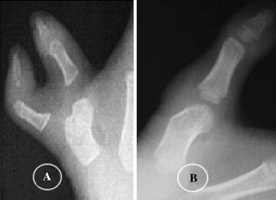
Materials and methods: A retrospective study was undertaken from January 1995 to December 2006. Clinical and radiographic evaluations were made according to Wassel's classification. The patients were performed by transferring an epiphyseal segment of the proximal phalanx with insertion of the abductor pollicis brevis tendon into the radial side of the epiphyseal proximal phalanx of the ulnar thumb. All patients were operated using one of five surgical procedures for bicephalous metacarpus, cartilaginous connection between the radial and ulnar proximal phalanges, the angular deformity of the metacarpophalangeal joint (MPJ) is >20°, and zigzag deformities. The postoperative results of the patients were evaluated for both function and cosmesis according to Tien's modified Tada scoring system.
Results: One hundred and sixty-four patients (102 females, 62 males) were included in this study. The MPJ was stable in 170 thumbs, 15 thumbs had 10° of radial instability, and new collateral ligaments were augmented in 27 thumbs. The alignment was normal in 75 thumbs, with alignment of the interphalangeal joint (IPJ) in 101 thumbs and alignment of the MPJ in 75 thumbs. Postoperatively, there were zigzag deformities in four thumbs (developed zigzag in two thumbs, recurrent zigzag in two thumbs); there was no first web space in those hands. There were four of 185 thumbs with thumb stiffness. The abductor function of 185 thumbs was as follows: >70° in 158 thumbs (85.4%), 50°-70° in 21 thumbs (11.4%), and <50° in six thumbs (3.2%). At the latest follow-up evaluation, no evidence of physeal growth injury or growth arrest was observed in any patient. Overall, we attained good results in 140 thumbs (75.7%), fair results in 36 thumbs (19.4%), and poor results in nine thumbs (4.9%).
Conclusion: We recommend the use of an epiphyseal segment of the proximal phalanx with insertion of the abductor pollicis brevis tendon into the radial side of the epiphyseal proximal phalanx of the ulnar thumb and to restore anatomical insertion of the abductor pollicis brevis muscle. The technique is simple, safe, and effective for thumb abductor function in the treatment of bifid thumb type IV in children.
Keywords: Congenital duplication; Duplicated thumb; Polydactyly; Thumb.
Figures


Similar articles
-
[CLINICAL RESEARCH OF POSTOPERATIVE DEVIATION SECONDARY TO THUMB DUPLICATION RESECTION].Zhongguo Xiu Fu Chong Jian Wai Ke Za Zhi. 2014 Jul;28(7):835-9. Zhongguo Xiu Fu Chong Jian Wai Ke Za Zhi. 2014. PMID: 26462345 Chinese.
-
Radial collateral ligament repair of the thumb metacarpophalangeal joint using the abductor pollicis brevis tendon.Plast Reconstr Surg. 2006 Feb;117(2):491-6. doi: 10.1097/01.prs.0000197219.77994.b2. Plast Reconstr Surg. 2006. PMID: 16462331
-
Opponensplasty With Abductor Pollicis Brevis Rerouting for Types II and IIIA Hypoplastic Thumbs.J Hand Surg Am. 2025 Jun;50(6):749.e1-749.e6. doi: 10.1016/j.jhsa.2024.02.006. Epub 2024 Apr 5. J Hand Surg Am. 2025. PMID: 38583166
-
[Extensor tendon injuries of the thumb].Unfallchirurg. 2021 Apr;124(4):287-293. doi: 10.1007/s00113-021-00982-z. Epub 2021 Mar 3. Unfallchirurg. 2021. PMID: 33656563 Review. German.
-
Instability of the metacarpophalangeal joint of the thumb. Reconstruction of the collateral ligaments using the extensor pollicis brevis tendon.J Bone Joint Surg Am. 1976 Jan;58(1):106-12. J Bone Joint Surg Am. 1976. PMID: 765344 Review.
Cited by
-
Resection and reconstruction for radial polydactyly Type IV-D in 206 cases: a retrospective clinical analysis.BMC Musculoskelet Disord. 2022 Feb 22;23(1):167. doi: 10.1186/s12891-022-05119-w. BMC Musculoskelet Disord. 2022. PMID: 35193542 Free PMC article.
-
Aesthetic outcomes of and anatomic reconstruction for Wassel type IV-D radial polydactyly using a modified Bilhaut-Cloquet procedure.Front Pediatr. 2023 Jul 7;11:1192168. doi: 10.3389/fped.2023.1192168. eCollection 2023. Front Pediatr. 2023. PMID: 37484763 Free PMC article.
-
[Recent progress in research of congenital polydactyly].Zhongguo Xiu Fu Chong Jian Wai Ke Za Zhi. 2018 Jul 15;32(7):827-831. doi: 10.7507/1002-1892.201806091. Zhongguo Xiu Fu Chong Jian Wai Ke Za Zhi. 2018. PMID: 30129303 Free PMC article. Chinese.
References
LinkOut - more resources
Full Text Sources
