

Spontaneous brain activity observed with functional magnetic resonance imaging as a potential biomarker in neuropsychiatric disorders
- PMID: 22132039
- PMCID: PMC2974101
- DOI: 10.1007/s11571-010-9126-9
Spontaneous brain activity observed with functional magnetic resonance imaging as a potential biomarker in neuropsychiatric disorders
Abstract
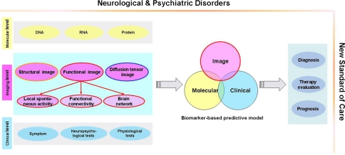
As functional magnetic resonance imaging (fMRI) studies have yielded increasing amounts of information about the brain's spontaneous activity, they have revealed fMRI's potential to locate changes in brain hemodynamics that are associated with neuropsychiatric disorders. In this paper, we review studies that support the notion that changes in brain spontaneous activity observed by fMRI can be used as potential biomarkers for diagnosis and treatment evaluation in neuropsychiatric disorders. We first review the methods used to study spontaneous activity from the perspectives of (1) the properties of local spontaneous activity, (2) the spatial pattern of spontaneous activity, and (3) the topological properties of brain networks. We also summarize the major findings associated with major neuropsychiatric disorders obtained using these methods. Then we review the pilot studies that have used spontaneous activity to discriminate patients from normal controls. Finally, we discuss current challenges and potential research directions to further elucidate the clinical use of spontaneous brain activity in neuropsychiatric disorders.
Keywords: Alzheimer’s disease; Functional connectivity; Low frequency fluctuation; Resting-state fMRI; Schizophrenia.
Figures
Similar articles
-
Clinical utility of resting-state functional connectivity magnetic resonance imaging for mood and cognitive disorders.J Neural Transm (Vienna). 2017 Jul;124(7):821-839. doi: 10.1007/s00702-017-1710-2. Epub 2017 Mar 23. J Neural Transm (Vienna). 2017. PMID: 28337552 Review.
-
Abnormal amplitude of low frequency fluctuation and functional connectivity in non-neuropsychiatric systemic lupus erythematosus: a resting-state fMRI study.Neuroradiology. 2019 Mar;61(3):331-340. doi: 10.1007/s00234-018-2138-6. Epub 2019 Jan 12. Neuroradiology. 2019. PMID: 30637462
-
Resting-state fMRI as a biomarker for Alzheimer's disease?Alzheimers Res Ther. 2012 Mar 15;4(2):8. doi: 10.1186/alzrt106. Alzheimers Res Ther. 2012. PMID: 22423634 Free PMC article.
-
Functional magnetic resonance imaging: basic principles and application in the neurosciences.Radiologia (Engl Ed). 2018 Sep-Oct;60(5):368-377. doi: 10.1016/j.rx.2017.12.007. Epub 2018 Mar 12. Radiologia (Engl Ed). 2018. PMID: 29544987 English, Spanish.
-
How do abnormalities in the brain's spontaneous activity translate into symptoms in schizophrenia? From an overview of resting state activity findings to a proposed spatiotemporal psychopathology.Prog Neurobiol. 2016 Oct-Nov;145-146:26-45. doi: 10.1016/j.pneurobio.2016.08.003. Epub 2016 Aug 12. Prog Neurobiol. 2016. PMID: 27531135 Review.
Cited by
-
Long genes are more frequently affected by somatic mutations and show reduced expression in Alzheimer's disease: Implications for disease etiology.Alzheimers Dement. 2021 Mar;17(3):489-499. doi: 10.1002/alz.12211. Epub 2020 Oct 19. Alzheimers Dement. 2021. PMID: 33075204 Free PMC article.
-
Intervention-induced enhancement in intrinsic brain activity in healthy older adults.Sci Rep. 2014 Dec 4;4:7309. doi: 10.1038/srep07309. Sci Rep. 2014. PMID: 25472002 Free PMC article. Clinical Trial.
-
Controllability in attention deficit hyperactivity disorder brains.Cogn Neurodyn. 2024 Aug;18(4):2003-2013. doi: 10.1007/s11571-023-10063-z. Epub 2024 Feb 6. Cogn Neurodyn. 2024. PMID: 39104674 Free PMC article.
-
Power spectral density and coherence analysis of Alzheimer's EEG.Cogn Neurodyn. 2015 Jun;9(3):291-304. doi: 10.1007/s11571-014-9325-x. Epub 2014 Dec 16. Cogn Neurodyn. 2015. PMID: 25972978 Free PMC article.
-
Potential correlations between abnormal homogeneity of default mode network and personality or lipid level in major depressive disorder.Brain Behav. 2024 Jul;14(7):e3622. doi: 10.1002/brb3.3622. Brain Behav. 2024. PMID: 39021241 Free PMC article.
References
-
- Allen G, Barnard H, McColl R, Hester AL, Fields JA, Weiner MF, Ringe WK, Lipton AM, Brooker M, McDonald E, Rubin CD, Cullum CM. Reduced hippocampal functional connectivity in Alzheimer disease. Arch Neurol. 2007;64:1482–1487. - PubMed
-
- Arthurs OJ, Boniface S. How well do we understand the neural origins of the fMRI BOLD signal? Trends Neurosci. 2002;25:27–31. - PubMed
LinkOut - more resources
Full Text Sources
Research Materials
