

Reactivation of M. tuberculosis infection in trans-membrane tumour necrosis factor mice
- PMID: 22132068
- PMCID: PMC3221652
- DOI: 10.1371/journal.pone.0025121
Reactivation of M. tuberculosis infection in trans-membrane tumour necrosis factor mice
Abstract
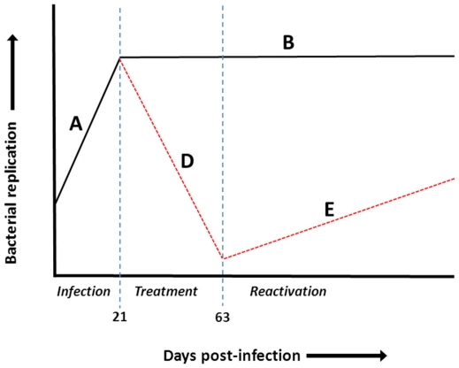
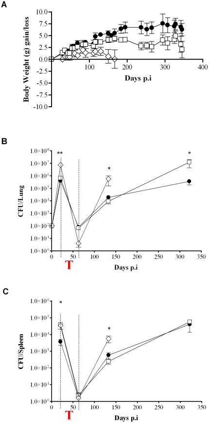
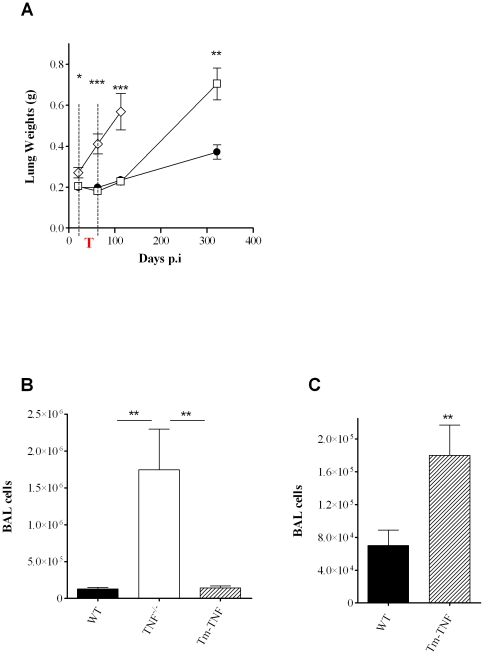
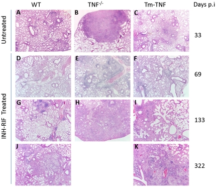
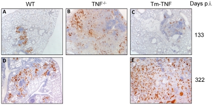
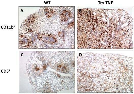
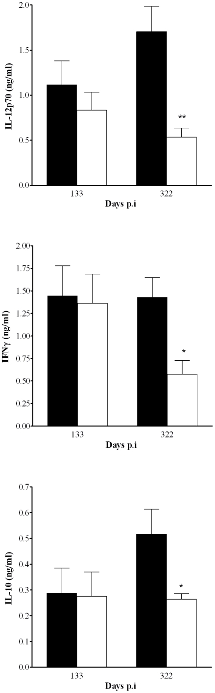
Of those individuals who are infected with M. tuberculosis, 90% do not develop active disease and represents a large reservoir of M. tuberculosis with the potential for reactivation of infection. Sustained TNF expression is required for containment of persistent infection and TNF neutralization leads to tuberculosis reactivation. In this study, we investigated the contribution of soluble TNF (solTNF) and transmembrane TNF (Tm-TNF) in immune responses generated against reactivating tuberculosis. In a chemotherapy induced tuberculosis reactivation model, mice were challenged by aerosol inhalation infection with low dose M. tuberculosis for three weeks to establish infection followed chemotherapeutic treatment for six weeks, after which therapy was terminated and tuberculosis reactivation investigated. We demonstrate that complete absence of TNF results in host susceptibility to M. tuberculosis reactivation in the presence of established mycobacteria-specific adaptive immunity with mice displaying unrestricted bacilli growth and diffused granuloma structures compared to WT control mice. Interestingly, bacterial re-emergence is contained in Tm-TNF mice during the initial phases of tuberculosis reactivation, indicating that Tm-TNF sustains immune pressure as in WT mice. However, Tm-TNF mice show susceptibility to long term M. tuberculosis reactivation associated with uncontrolled influx of leukocytes in the lungs and reduced IL-12p70, IFNγ and IL-10, enlarged granuloma structures, and failure to contain mycobacterial replication relative to WT mice. In conclusion, we demonstrate that both solTNF and Tm-TNF are required for maintaining immune pressure to contain reactivating M. tuberculosis bacilli even after mycobacteria-specific immunity has been established.
Conflict of interest statement
Figures
Similar articles
-
Efficacy of membrane TNF mediated host resistance is dependent on mycobacterial virulence.Tuberculosis (Edinb). 2008 May;88(3):221-34. doi: 10.1016/j.tube.2007.08.011. Epub 2007 Oct 22. Tuberculosis (Edinb). 2008. PMID: 17950671
-
Reactivation of latent tuberculosis by an inhibitor of inducible nitric oxide synthase in an aerosol murine model.Immunology. 2002 Nov;107(3):350-7. doi: 10.1046/j.1365-2567.2002.01511.x. Immunology. 2002. PMID: 12423311 Free PMC article.
-
Tumor necrosis factor blockade in chronic murine tuberculosis enhances granulomatous inflammation and disorganizes granulomas in the lungs.Infect Immun. 2008 Mar;76(3):916-26. doi: 10.1128/IAI.01011-07. Epub 2008 Jan 22. Infect Immun. 2008. PMID: 18212087 Free PMC article.
-
Reactivation of tuberculosis by tumor necrosis factor neutralization.Eur Cytokine Netw. 2007 Mar;18(1):5-13. doi: 10.1684/ecn.2007.0083. Epub 2007 Mar 30. Eur Cytokine Netw. 2007. PMID: 17400533 Review.
-
Role of Tumor Necrosis Factor in Tuberculosis.Biomolecules. 2025 May 12;15(5):709. doi: 10.3390/biom15050709. Biomolecules. 2025. PMID: 40427602 Free PMC article. Review.
Cited by
-
The contraceptive depot medroxyprogesterone acetate impairs mycobacterial control and inhibits cytokine secretion in mice infected with Mycobacterium tuberculosis.Infect Immun. 2013 Apr;81(4):1234-44. doi: 10.1128/IAI.01189-12. Epub 2013 Feb 4. Infect Immun. 2013. PMID: 23381991 Free PMC article.
-
TNFRp75-dependent immune regulation of alveolar macrophages and neutrophils during early Mycobacterium tuberculosis and Mycobacterium bovis BCG infection.Immunology. 2021 Feb;162(2):220-234. doi: 10.1111/imm.13277. Epub 2020 Oct 23. Immunology. 2021. PMID: 33020922 Free PMC article.
-
Mycobacterium tuberculosis Rv0341 Promotes Mycobacterium Survival in In Vitro Hostile Environments and within Macrophages and Induces Cytokines Expression.Pathogens. 2020 Jun 8;9(6):454. doi: 10.3390/pathogens9060454. Pathogens. 2020. PMID: 32521796 Free PMC article.
-
Transmembrane TNF and Its Receptors TNFR1 and TNFR2 in Mycobacterial Infections.Int J Mol Sci. 2021 May 22;22(11):5461. doi: 10.3390/ijms22115461. Int J Mol Sci. 2021. PMID: 34067256 Free PMC article. Review.
-
Tumor necrosis factor neutralization combined with chemotherapy enhances Mycobacterium tuberculosis clearance and reduces lung pathology.Am J Clin Exp Immunol. 2013 Feb 27;2(1):124-34. Print 2013. Am J Clin Exp Immunol. 2013. PMID: 23885330 Free PMC article.
References
-
- WHO. 2010. GLOBAL TURCULOSIS CONTROL.
-
- Van Rhijn I, Godfroid J, Michel A, Rutten V. Bovine tuberculosis as a model for human tuberculosis: advantages over small animal models. Microbes Infect. 2008;10:711–715. - PubMed
Publication types
MeSH terms
Substances
LinkOut - more resources
Full Text Sources
Medical
Molecular Biology Databases
