Shapes of discoid intracellular compartments with small relative volumes
- PMID: 22132079
- PMCID: PMC3221666
- DOI: 10.1371/journal.pone.0026824
Shapes of discoid intracellular compartments with small relative volumes
Abstract
A prominent feature of many intracellular compartments is a large membrane surface area relative to their luminal volume, i.e., the small relative volume. In this study we present a theoretical analysis of discoid membrane compartments with a small relative volume and then compare the theoretical results to quantitative morphological assessment of fusiform vesicles in urinary bladder umbrella cells. Specifically, we employ three established extensions of the standard approach to lipid membrane shape calculation and determine the shapes that could be expected according to three scenarios of membrane shaping: membrane adhesion in the central discoid part, curvature driven lateral segregation of membrane constituents, and existence of stiffer membrane regions, e.g., support by protein scaffolds. The main characteristics of each scenario are analyzed. The results indicate that even though all three scenarios can lead to similar shapes, there are values of model parameters that yield qualitatively distinctive shapes. Consequently, a distinctive shape of an intracellular compartment may reveal its membrane shaping mechanism and the membrane structure. The observed shapes of fusiform vesicles fall into two qualitatively different classes, yet they are all consistent with the theoretical results and the current understanding of their structure and function.
Conflict of interest statement
Figures


 m (A), 100 nm (B–E).
 at a vanishing relative difference between the lateral tensions of the bilayer leaflets (
 ). At
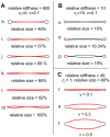
). At  , the shape is the well-known biconcave shape of a red blood cell (shape a). Decreasing the volume leads to a contact of the membrane at the discoid center (shape b,
  ) and then to an increase of the contact surface area (shape c,
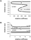
  ). B) Phase diagram describing the values of
  and
and  at which the membrane of a discoid compartment comes into contact with itself. The positions of the shapes a, b and c are presented.
 ; an increase of
  increases the contact surface area. The impact of the lateral segregation is described by parameter
  ; an increase in
  corresponds to more convex shapes with a smaller contact surface area. The relative volume of all shapes is 0.1.
 ), the spontaneous curvature (
 ) and the relative size of the stiffer regions, and the relative volume of the compartment (
 ) are denoted. The spontaneous curvature of the soft region is zero. A) Effects of an increasing size of a very stiff region with
  in the central discoid part. B) Effects of an increasing size of a curved (
 ) stiffer region supporting the discoid rim. C) Effects of an increasing relative volume in the case corresponding to the observed FV: the stiffer region is in the central discoid part, it occupies 60% of the total membrane surface area and has
  . The relative stiffness of the stiffer region is chosen to be 40.
 and
and  ). The crossed circles correspond to the shapes presented in Fig. 5.
Similar articles
-
Equilibrium shapes of erythrocytes in rouleau formation.Biophys J. 2003 Mar;84(3):1486-92. doi: 10.1016/S0006-3495(03)74961-3. Biophys J. 2003. PMID: 12609855 Free PMC article.
-
Quantifying immunogold labelling patterns of cellular compartments when they comprise mixtures of membranes (surface-occupying) and organelles (volume-occupying).Histochem Cell Biol. 2008 Mar;129(3):367-78. doi: 10.1007/s00418-007-0375-6. Epub 2008 Jan 5. Histochem Cell Biol. 2008. PMID: 18180944
-
Endoplasmatic reticulum shaping by generic mechanisms and protein-induced spontaneous curvature.Adv Colloid Interface Sci. 2014 Jun;208:153-60. doi: 10.1016/j.cis.2014.02.006. Epub 2014 Feb 15. Adv Colloid Interface Sci. 2014. PMID: 24607001 Review.
-
Trans-membrane area asymmetry controls the shape of cellular organelles.Int J Mol Sci. 2015 Mar 9;16(3):5299-333. doi: 10.3390/ijms16035299. Int J Mol Sci. 2015. PMID: 25761238 Free PMC article.
-
Compartmentation of the rough endoplasmic reticulum.Mol Cell Biochem. 1986 Jun;71(1):3-18. doi: 10.1007/BF00219323. Mol Cell Biochem. 1986. PMID: 2425244 Review.
Cited by
-
Gold nanoparticles as physiological markers of urine internalization into urothelial cells in vivo.Int J Nanomedicine. 2013;8:3945-53. doi: 10.2147/IJN.S44363. Epub 2013 Oct 14. Int J Nanomedicine. 2013. PMID: 24143099 Free PMC article.
-
Electron tomography of fusiform vesicles and their organization in urothelial cells.PLoS One. 2012;7(3):e32935. doi: 10.1371/journal.pone.0032935. Epub 2012 Mar 12. PLoS One. 2012. PMID: 22427911 Free PMC article.
-
Cryo-EM structures of peripherin-2 and ROM1 suggest multiple roles in photoreceptor membrane morphogenesis.Sci Adv. 2022 Nov 11;8(45):eadd3677. doi: 10.1126/sciadv.add3677. Epub 2022 Nov 9. Sci Adv. 2022. PMID: 36351012 Free PMC article.
-
Urothelial endocytic vesicle recycling and lysosomal degradative pathway regulated by lipid membrane composition.Histochem Cell Biol. 2013 Feb;139(2):249-65. doi: 10.1007/s00418-012-1034-0. Epub 2012 Oct 12. Histochem Cell Biol. 2013. PMID: 23064746
-
Membrane lipids and proteins as modulators of urothelial endocytic vesicles pathways.Histochem Cell Biol. 2013 Nov;140(5):507-20. doi: 10.1007/s00418-013-1095-8. Epub 2013 Apr 27. Histochem Cell Biol. 2013. PMID: 23624723 Review.
References
-
- Shibata Y, Hu J, Kozlov MM, Rapoport TA. Mechanisms shaping the membranes of cellular organelles. Annu Rev Cell Dev Biol. 2009;25:329–354. - PubMed
-
- McMahon HT, Gallop JL. Membrane curvature and mechanisms of dynamic cell membrane remodelling. Nature. 2005;438:590–596. - PubMed
-
- Zimmerberg J, Kozlov MM. How proteins produce cellular membrane curvature. Nat Rev Mol Cell Biol. 2005;7:9–19. - PubMed
-
- Seifert U. Configurations of uid membranes and vesicles. Adv Phys. 1997;46:13–137.
-
- Porter KR, Bonneville MA. An Introduction To The Fine Structure of Cells And Tissues. 1963. Lea & Febiger: Philadelphia.
Publication types
MeSH terms
LinkOut - more resources
Full Text Sources

