. 2011;6(11):e27583.
doi: 10.1371/journal.pone.0027583. Epub 2011 Nov 23.

Deficient spindle assembly checkpoint in multiple myeloma

Affiliations

Deficient spindle assembly checkpoint in multiple myeloma

Elena Díaz-Rodríguez et al. PLoS One. 2011.

Abstract

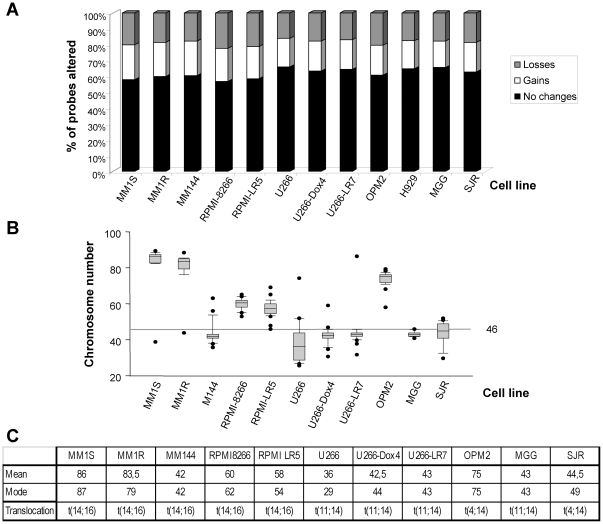
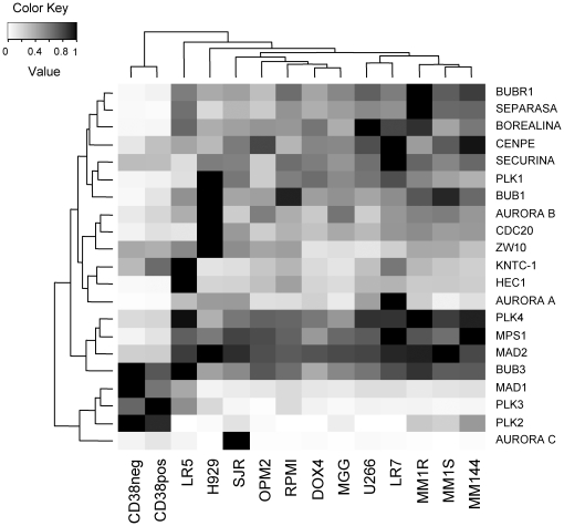
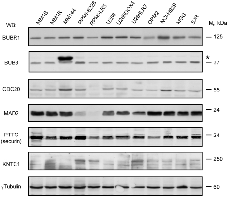
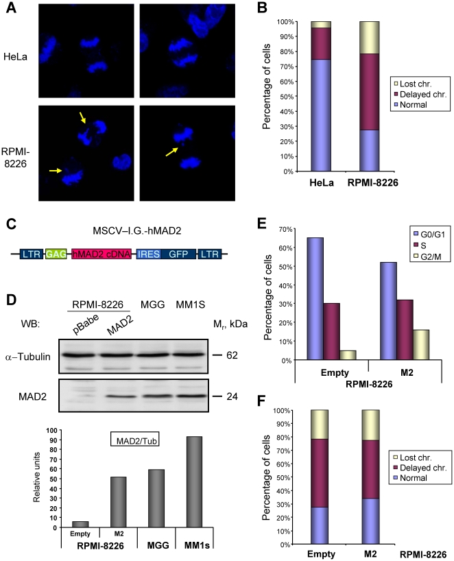
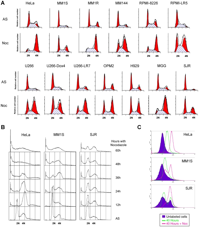
Multiple myeloma (MM) is a hematological disease characterized by an abnormal accumulation of plasma cells in the bone marrow. These cells have frequent cytogenetic abnormalities including translocations of the immunoglobulin heavy chain gene and chromosomal gains and losses. In fact, a singular characteristic differentiating MM from other hematological malignancies is the presence of a high degree of aneuploidies. As chromosomal abnormalities can be generated by alterations in the spindle assembly checkpoint (SAC), the functionality of such checkpoint was tested in MM. When SAC components were analyzed in MM cell lines, the RNA levels of most of them were conserved. Nevertheless, the protein content of some key constituents was very low in several cell lines, as was the case of MAD2 or CDC20 in RPMI-8226 or RPMI-LR5 cells. The recovery of their cellular content did not substantially affect cell growth, but improved their ability to segregate chromosomes. Finally, SAC functionality was tested by challenging cells with agents disrupting microtubule dynamics. Most of the cell lines analyzed exhibited functional defects in this checkpoint. Based on the data obtained, alterations both in SAC components and their functionality have been detected in MM, pointing to this pathway as a potential target in MM treatment.


Conflict of interest statement

Competing Interests: The authors have declared that no competing interests exist.

Figures

Figure 1
Figure 1. MM cell lines are genetically unstable and typically aneuploid.
A. Representation of the karyotype obtained by CGH arrays on genomic DNA of the indicated cell lines. The percentage of the total number of probes that are conserved, gained or lost are indicated in different colors (shown on the right). B. Representation of the karyopype obtained with conventional metaphase chromosome counts. C. Mean chromosome numbers as well as the mode of 50 metaphases counted in the indicated cell lines are shown. Besides, the characteristic translocation found on each cell line is detailed in the bottom row.
Figure 2
Figure 2. Expression of SAC proteins in MM cell lines.
Total RNA purified from the different MM cell lines as well as plasma cells (CD38pos) or other white blood cells (CD38neg) was converted into cDNA by the reverse transcriptase reaction using oligo-dT. Quantitative PCR reaction was performed with oligonucleotide pairs specific for the indicated proteins (indicated on the right) on the different cell lines (shown on the bottom). The relative amount of the different proteins were normalized with the housekeeping gene GAPDH and represented in a heat map.
Figure 3
Figure 3. Protein expression of different SAC components in MM.
Protein extracts from the MM cell lines indicated at the top of the figure were prepared, quantified, and 50 µg separated by SDS-PAGE. Once transferred, membranes were proved with antibodies specifically detecting the proteins shown on the left.
Figure 4
Figure 4. Characterization of the recovery of MAD2 levels on RPMI-8226 cells by retroviral transduction.
A. RPMI-8226 cells exhibit chromosome segregation defects. Hela or RPMI-8226 cells were plated on glass coverslips and 2 days later fixed, counterstained with DAPI and anaphases counted at a fluorescence microscope. Yellow arrows point to chromosomal defects. B. The presence of chromosomes left behind (delayed chromosomes) or lost between the two daughter cells was measured in at least 50 anaphases in the indicated cell lines and represented as the percentage of cells with the different defect. C. Schematic representation of the plasmid used to transduce MM cells with hMAD2. D-F. Recovery of MAD2 levels in the MM cell line RPMI-8226. RPMI-8226 cells were transduced with the vector shown in panel C or empty vector, and 2 days later plated for the subsequent analysis. D. Recovery of MAD2 protein levels. Protein extracts from the indicated cell lines were prepared, 30 µg separated by SDS-PAGE and MAD2 and α-tubulin amount determined by regular WB as indicated (upper panels). Another set was similarly analyzed, but their levels were quantified using appropriate secondary antibodies and the Odyssey software. The relative ratio of both proteins is shown in the lower graph. E. The recovery of MAD2 levels induces a small change in their cell cycle profile, producing the accumulation of more cells in the G2/M transition. F. Besides, a small improvement in chromosomal dynamics in mitosis is detected after MAD2 recovery, measured as described in B.
Figure 5
Figure 5. MM cell lines exhibit a deficient SAC when challenged with nocodazole.
A. Several MM cell lines were treated with nocodazole for 20 hours, and their cell cycle status after the arrest measured by conventional flow cytometry after DNA staining. Most of the MM cell lines analyzed was not as efficiently arrested in those conditions as HeLa cells. B. The indicated cell lines were arrested for up to 60 hours with nocodazole to determine if the deficient arrest was due to a slower proliferation. The increase in the time of treatment did not effectively synchronize MM1S or SJR cells. C. Cells were labeled for 30 minutes with the CellTracker Red dye, washed out the excess and placed in culture in normal medium (green line), or supplemented with nocodazole (pink line). Cell division in untreated cells made them loose staining, as indicated by the shift in the peak. When the arrest was efficient, the population did not shift (see HeLa cells). Nevertheless, part of the nocodazole treated MM1S population shifted as the control confirming that those cells had, in fact, divided even in the presence of nocodazole. On the other hand the low synchronization detected in SJR was mainly due to the low proliferation of this cell line given that even the normal population had not divided in 40 hours.

References

    1. Jemal A, Siegel R, Ward E, Murray T, Xu J, et al. Cancer statistics, 2007. CA Cancer J Clin. 2007;57:43–66. - PubMed
    1. Raab MS, Podar K, Breitkreutz I, Richardson PG, Anderson KC. Multiple myeloma. Lancet. 2009;374:324–339. - PubMed
    1. Ocio EM, Mateos MV, Maiso P, Pandiella A, San-Miguel JF. New drugs in multiple myeloma: mechanisms of action and phase I/II clinical findings. Lancet Oncol. 2008;9:1157–1165. - PubMed
    1. Gutierrez NC, Garcia-Sanz R, San Miguel JF. Molecular biology of myeloma. Clin Transl Oncol. 2007;9:618–624. - PubMed
    1. Kuehl WM, Bergsagel PL. Multiple myeloma: evolving genetic events and host interactions. Nat Rev Cancer. 2002;2:175–187. - PubMed

Publication types

MeSH terms