. 2011;6(11):e27881.
doi: 10.1371/journal.pone.0027881. Epub 2011 Nov 21.

Astrocyte proliferation following stroke in the mouse depends on distance from the infarct

Affiliations

Astrocyte proliferation following stroke in the mouse depends on distance from the infarct

George E Barreto et al. PLoS One. 2011.

Abstract

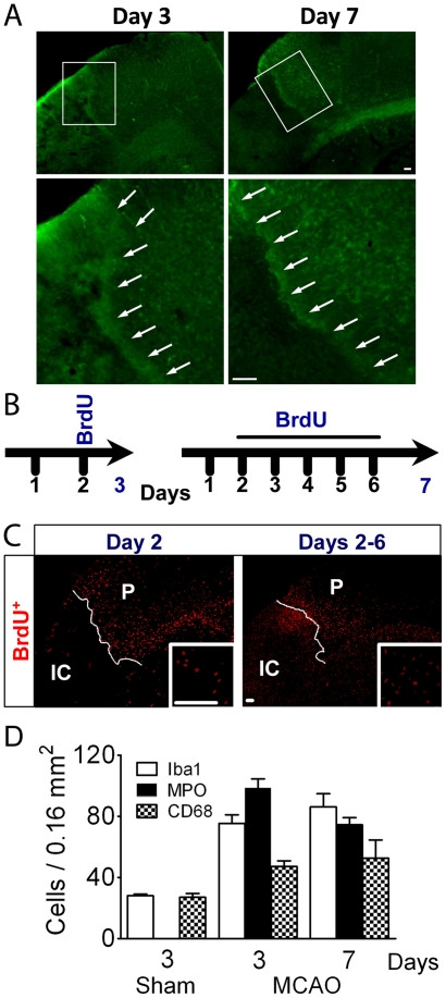
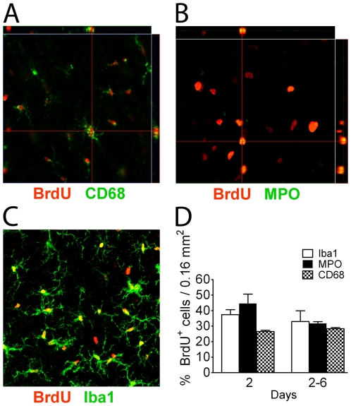
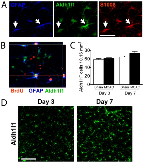
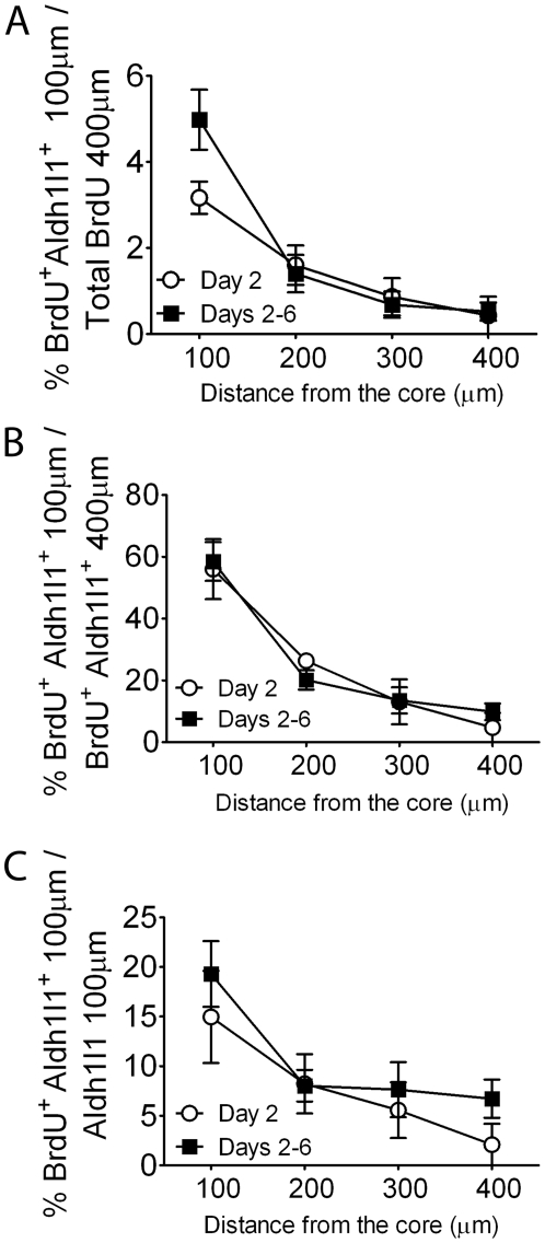
Reactive gliosis is a hallmark of brain pathology and the injury response, yet the extent to which astrocytes proliferate, and whether this is central to astrogliosis is still controversial. We determined the fraction of mature astrocytes that proliferate in a mouse stroke model using unbiased stereology as a function of distance from the infarct edge. Cumulatively 11.1±1.2% of Aldh1l1(+) astrocytes within 400 µm in the cortical penumbra incorporate BrdU in the first week following stroke, while the overall number of astrocytes does not change. The number of astrocytes proliferating fell sharply with distance with more than half of all proliferating astrocytes found within 100 µm of the edge of the infarct. Despite extensive cell proliferation primarily of microglia and neutrophils/monocytes in the week following stroke, few mature astrocytes re-enter cell cycle, and these are concentrated close to the infarct boundary.


Conflict of interest statement

Competing Interests: The authors have declared that no competing interests exist.

Figures

Figure 1
Figure 1. Cellular proliferation assessed with BrdU.
(A) Low power micrographs show the edge of the core outlined by Aldh1l1 fluorescent astrocytes (green) present at the boundary visibly separating the core from the surrounding tissue on days 3 and 7 after ischemia; higher magnification of the areas indicated in the white boxes are shown below the low power images. (B) To study the proliferative response BrdU was injected 3 times/day on day 2 and animals were sacrificed on day 3 to determine the early response. A second group was injected 3 times/day from days 2–6 to analyze cumulative proliferation over the week following stroke, with animals sacrificed on day 7. (C) Panoramic views of sections immunostained for BrdU show proliferation in both penumbra (P) and ischemic core (IC), with somewhat stronger staining in penumbra on day 2, while the core showed more labelled cells on days 2–6. (D) Morphometric analysis shows the density of each cell type in the penumbra, demonstrating an increased inflammatory reaction surrounding the core. Sham animals were also analyzed for the number of each cell type on day 3 and 7, but the numbers for each cell type did not change significantly, so only the data for day 3 is shown. No MPO positive cells were detected in the sham animals. Scale Bar, 50 µm.
Figure 2
Figure 2. Analysis of proliferating cells in the penumbra identifies many inflammatory cells.
Using different antibodies to identify the cell types labelled with BrdU, reveals that most proliferating cells are microglia/macrophages (A, C) and monocytes/neutrophils by colocalization (B) of cell type markers with BrdU, and this was confirmed by the morphometric analysis (D). N = 3 animals for day 2, N = 5 animals for day 7. Scale Bar, 50 µm.
Figure 3
Figure 3. Few Aldh1l1+ astrocytes proliferate in the cortical penumbra.
(A) Triple labelling reveals that most astrocytes in the cortical penumbra label for GFAP, Aldh1l1 and S100β. (B) Orthogonal view of a mature astrocyte co-expressing Aldh1l1, BrdU and GFAP in the cortical penumbra. (C) The number of Aldh1l1+ astrocytes assessed in layers II–III of the cortical penumbra does not change over the first week post-injury, or compared to sham animals. (D) Illustrative confocal micrographs show increased prominence of Aldh1l1 positive cells with cellular hypertrophy, but no change in overall number. Scale Bar, 50 µm.
Figure 4
Figure 4. Detection of apoptotic cells in the penumbra.
(A) Apoptotic cells identified by staining for cleaved caspase 3 show few apoptotic cells in the core and some in the penumbra on day 3, contrasting with a striking increase in apoptotic cells in the core on day 7. (B) Quantitation of apoptotic cells in the penumbra on days 3 and 7 after injury. (C) A small percent of Aldh1l1 cells are also apoptotic as assessed by caspase 3 activation. The number significantly decreases between days 3 and 7. IC = ischemic core; P = penumbra. N = 3 mice for day 2, N = 5 mice for day 7. Scale Bar, 50 µm.
Figure 5
Figure 5. Astrocyte proliferation as a function of distance from the infarct core.
BrdU labelling on day 2 or 2–6 cumulatively shows a small percentage of mature Aldh1l1+ astrocytes proliferate after MCAO within 400 µm of the ischemic core. Stereological analysis of astrocyte proliferation as a function of distance shows that Aldh1l1+ cells mainly co-express BrdU in the first 200 µm from the infarct area; the number decreases dramatically with distance. The fraction of proliferating astrocytes compared to all proliferating cells in the penumbra is low (A), and the fraction of proliferating astrocytes present falls rapidly with distance (B), and the fraction of all astrocytes present that proliferate is highest close to the core (C).

References

    1. Pekny M, Nilsson M. Astrocyte activation and reactive gliosis. Glia. 2005;50:427–434. - PubMed
    1. Buffo A, Rite I, Tripathi P, Lepier A, Colak D, et al. Origin and progeny of reactive gliosis: A source of multipotent cells in the injured brain. Proc Natl Acad Sci U S A. 2008;105:3581–3586. - PMC - PubMed
    1. Sofroniew MV. Molecular dissection of reactive astrogliosis and glial scar formation. Trends Neurosci. 2009;32:638–647. - PMC - PubMed
    1. Sofroniew MV, Vinters HV. Astrocytes: biology and pathology. Acta Neuropathol. 2010;119:7–35. - PMC - PubMed
    1. Cavanagh JB. The proliferation of astrocytes around a needle wound in the rat brain. J Anat. 1970;106:471–487. - PMC - PubMed

Publication types

Substances

LinkOut - more resources