. 2011;6(11):e27943.
doi: 10.1371/journal.pone.0027943. Epub 2011 Nov 21.

Phenotypes of non-attached Pseudomonas aeruginosa aggregates resemble surface attached biofilm

Affiliations

Phenotypes of non-attached Pseudomonas aeruginosa aggregates resemble surface attached biofilm

Morten Alhede et al. PLoS One. 2011.

Abstract

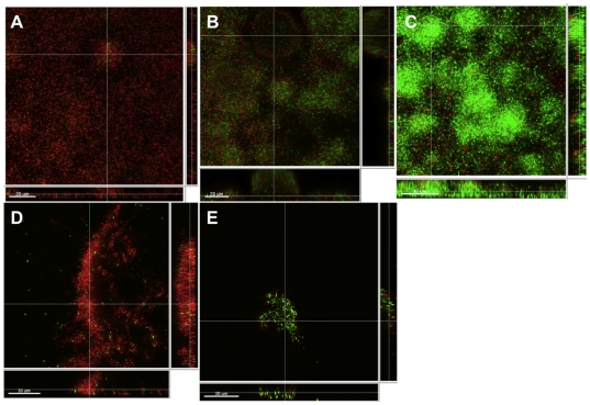
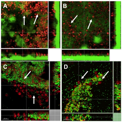
For a chronic infection to be established, bacteria must be able to cope with hostile conditions such as low iron levels, oxidative stress, and clearance by the host defense, as well as antibiotic treatment. It is generally accepted that biofilm formation facilitates tolerance to these adverse conditions. However, microscopic investigations of samples isolated from sites of chronic infections seem to suggest that some bacteria do not need to be attached to surfaces in order to establish chronic infections. In this study we employed scanning electron microscopy, confocal laser scanning microscopy, RT-PCR as well as traditional culturing techniques to study the properties of Pseudomonas aeruginosa aggregates. We found that non-attached aggregates from stationary-phase cultures have comparable growth rates to surface attached biofilms. The growth rate estimations indicated that, independently of age, both aggregates and flow-cell biofilm had the same slow growth rate as a stationary phase shaking cultures. Internal structures of the aggregates matrix components and their capacity to survive otherwise lethal treatments with antibiotics (referred to as tolerance) and resistance to phagocytes were also found to be strikingly similar to flow-cell biofilms. Our data indicate that the tolerance of both biofilms and non-attached aggregates towards antibiotics is reversible by physical disruption. We provide evidence that the antibiotic tolerance is likely to be dependent on both the physiological states of the aggregates and particular matrix components. Bacterial surface-attachment and subsequent biofilm formation are considered hallmarks of the capacity of microbes to cause persistent infections. We have observed non-attached aggregates in the lungs of cystic fibrosis patients; otitis media; soft tissue fillers and non-healing wounds, and we propose that aggregated cells exhibit enhanced survival in the hostile host environment, compared with non-aggregated bacterial populations.


Conflict of interest statement

Competing Interests: The authors have declared that no competing interests exist.

Figures

Figure 1
Figure 1. SEM survey of different P. aeruginosa cultures.
SEM of A – Aggregate harvested from a 48-h old stationary culture. B – Details of a 3-day old biofilm grown in flow-cell. C – Details of 48-h old stationary aggregate. D – Planktonic cells (OD600 = 0.5). Note that the single planktonic cells are difficult to fixate on the specimen during SEM preparation due to the small size (leads to few cells on the specimen).
Figure 2
Figure 2. Invstigation of the clonal relationship in clumps.
Mixed Yellow (YFP) and Cyan (CFP) constitually tagged PAO1 were inoculated together. The CLSM visualization demonstrated that the clumps consisted of both yellow and blue cells indicating non-clonal growth. Length of size bar: 6 µm.
Figure 3
Figure 3. Antibiotic tolerance of maturing flow-cell biofilms of P. aeruginosa and aggregates harvested from a static P. aeruginosa culture.
Biofilms and aggregates were grown for 24 h to 72-h prior to tobramycin (100 ug/ml) treatment for 24-h. For visualization by CLSM a GFP-tagged PAO1 strain (green) was used and stained with the DNA stain PI (red) for visualizing dead bacteria. Panels A, B and C represent biofilm tolerance on day 1, 2 and 3 respectively andpanels D and E are aggregates on day 1 and 2. Length of size bars: 20 µm.
Figure 4
Figure 4. Growth rates of flow-cell biofilms and aggregates over time.
Growth rates were estimated by quantifying rRNA molecules per rDNA molecules by RT-PCR.
Figure 5
Figure 5. Antibiotic tolerance of 48-h old aggregates harvested from a static P. aeruginosa culture and the effect of disruption by whirly mixing.
Aggregates were grown for 48-h prior to tobramycin (100 ug/ml) treatment for 24-h or whirly mix (20 seconds) followed by tobramycin treatment for 24 h. 24 h. Bars represent the median. Mann-Whitney test were chosen to compare the medians of the different treatment regimens. ns indicates no significant differences between treatments (p>0.05). * Indicates significant difference (p<0.05). *** Indicates a very significant difference (p<0.0001).
Figure 6
Figure 6. Effect of prolonged whirly mix.
Aggregates were grown for 48-h prior to tobramycin (100 ug/ml) treatment for 24-h, followed by whirly mix and another round of tobramycin treatment. Number indicates seconds of whirly mix. Bars represent the median. Kruskal-Wallis with Dunn's post test was chosen to compare the effect of whirly mix and subsequent treatment with tobramycin. The post test found the increased killing to be significant (P<0.05 when compared to 0 seconds) after 30 seconds of whirly mix.
Figure 7
Figure 7. Tolerance towards PMNs.
Flow-cell biofilms (A+B) were grown for 72-h before addition of PMNs and the aggregates (C+D) were grown for 48-h before the addition of PMNs. The images shows that aggregates are not phagocytosed or penetrated by PMNs. For visualization a GFP-tagged PAO1 strain (green) was used and SYTO62 was used to stain the PMNs (red). Arrows point at paralyzed PMNs. Length of size bars: 20 µm.
Figure 8
Figure 8. DNA content in P. aeruginosa cultures.
DNA (PI) staining of A - Aggregate harvested from a 48-h old stationary culture stained with PI. B – 3 day old biofilm grown in flow-cell stained with PI. C – GFP-tagged planktonic cells (OD – 0.5) stained with PI. Length of size bar: 15 µm.
Figure 9
Figure 9. Effect of DNase on aggregate formation.
Stationary cultures grown with 90 U/ml DNase I in the medium. After 48-h there was a clear difference of the visible aggregation in the culture. The control was left untreated. The top and bottom panels represent two independent experiments.
Figure 10
Figure 10. Matrix production by mutants.
We tested whether if mutants not able to produce the two distinct extracellular polysaccharides (Pel and Psl) could form intercellular fibers and if these fibers could be removed by DNAse treatment. WT - wild type PAO1, ΔpelA – PAO1 not able to produce Pel polysaccarides, ΔpslBCD - PAO1 not able to produce Psl polysaccarides, ΔpslBCDpelA - PAO1 not able to produce both Psl and Pel polysaccarides.
Figure 11
Figure 11. Localization of lections and DNA in aggregates.
HHA-FITC and PI staining of aggregate harvested from a 48-h old stationary culture. Figure 1 and 2 represent two independent experiments with A, B and C representing HHA-FITC, PI and HHA-FITC+PI. To test whether if the fibers are made from more than one polymer, we co-stained the aggregates with PI (red) and a mannose-specific lectin stain (HHA-FITC) to visualize DNA and any present Psl polymers. Length of size bar; Figure 1: 5 µm; Figure 2: 10 µm.

References

    1. Donlan RM. Biofilms: microbial life on surfaces. Emerg Infect Dis. 2002;8:881–890. - PMC - PubMed
    1. Høiby N, Bjarnsholt T, Givskov M, Molin S, Ciofu O. Antibiotic resistance of bacterial biofilms. Int J Antimicrob Agents. 2010;35:322–332. - PubMed
    1. Donlan RM, Costerton JW. Biofilms: survival mechanisms of clinically relevant microorganisms. Clin Microbiol Rev. 2002;15:167–193. - PMC - PubMed
    1. Costerton JW, Stewart PS, Greenberg EP. Bacterial biofilms: a common cause of persistent infections. Science. 1999;284:1318–1322. - PubMed
    1. Moreau-Marquis S, Stanton BA, O'Toole GA. Pseudomonas aeruginosa biofilm formation in the cystic fibrosis airway. Pulm Pharmacol Ther. 2008;21:595–599. - PMC - PubMed

Publication types

MeSH terms