

Experience with the use of prebent plates for the reconstruction of mandibular defects
- PMID: 22132258
- PMCID: PMC3052711
- DOI: 10.1055/s-0030-1268520
Experience with the use of prebent plates for the reconstruction of mandibular defects
Abstract
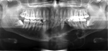
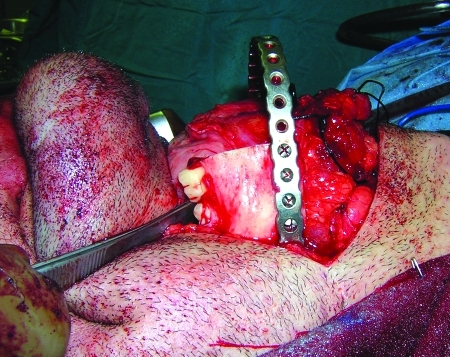
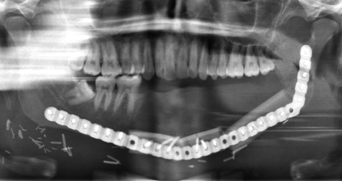
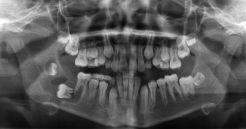
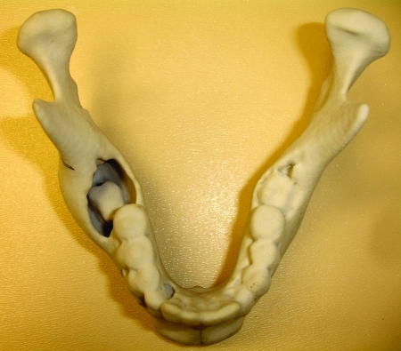
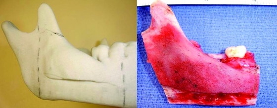
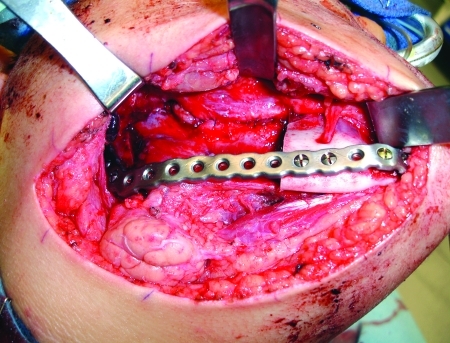
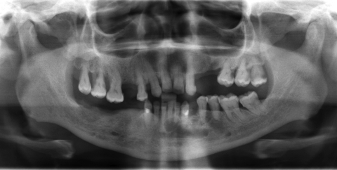
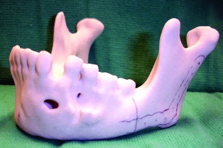
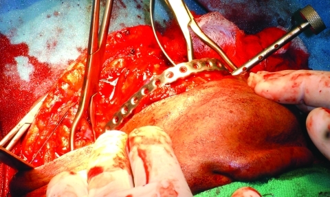
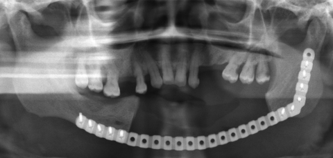
Bending of large titanium plates for mandibular reconstruction is a tedious task. This is usually done by trial and error over an intraoperatively bent template. By means of rapid prototype technology, accurate three-dimensional models can be obtained. Using these models, it is possible to design, obtain, and adapt custom hardware for individual surgical cases. Reductions of operating room time when using this technology have been reported from 17% to 60%, with an average of 20%. This translates to reduction of cost and risks, improving the overall surgical outcome. The purpose of this article is to establish the indications and contraindication for the use three-dimensional models and prebent plates. We present our experience with five cases in which prebent reconstruction plates were used for mandibular reconstruction. No significant complications occurred, and satisfactory results were achieved in all cases. We found that the models required to obtain the hardware are extremely accurate, have multiple reported applications, and represent a valuable surgical tool in the planning and execution of reconstructive surgery.
Keywords: Prebent plates; custom hardware; mandible reconstruction; medical rapid prototyping; stereolithographic models.
Figures
References
-
- Marx R E, Stern D. Oral and Maxillofacial Pathology: A Rationale for Diagnosis and Treatment. Carol Stream, IL: Quintessence Publishing Co., Inc.; 2003. pp. 635–703.
-
- Mark R E, Stevens M R. Atlas of Oral and Extraoral Bone Harvesting. Carol Stream, IL: Quintessence Publishing Co.; 2010. pp. 1–6.
-
- Marx R E. Bone and bone graft healing. Oral Maxillofac Surg Clin North Am. 2007;19:455–466, v. - PubMed
-
- Winder J, Bibb R. Medical rapid prototyping technologies: state of the art and current limitations for application in oral and maxillofacial surgery. J Oral Maxillofac Surg. 2005;63:1006–1015. - PubMed
-
- Mankovich N J, Cheeseman A M, Stoker N G. The display of three-dimensional anatomy with stereolithographic models. J Digit Imaging. 1990;3:200–203. - PubMed
LinkOut - more resources
Full Text Sources