. 2011 Dec 1:12:61.
doi: 10.1186/1471-2091-12-61.

Assembly and proteolytic processing of mycobacterial ClpP1 and ClpP2

Affiliations

Assembly and proteolytic processing of mycobacterial ClpP1 and ClpP2

Nadia Benaroudj et al. BMC Biochem. .

Abstract

Background: Caseinolytic proteases (ClpPs) are barrel-shaped self-compartmentalized peptidases involved in eliminating damaged or short-lived regulatory proteins. The Mycobacterium tuberculosis (MTB) genome contains two genes coding for putative ClpPs, ClpP1 and ClpP2 respectively, that are likely to play a role in the virulence of the bacterium.

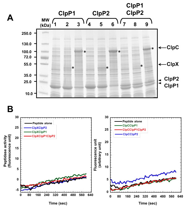
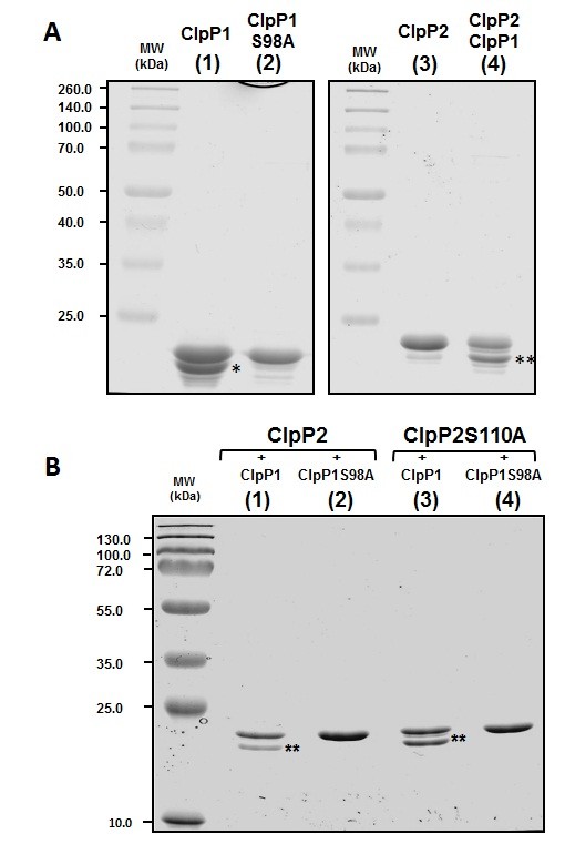
Results: We report the first biochemical characterization of ClpP1 and ClpP2 peptidases from MTB. Both proteins were produced and purified in Escherichia coli. Use of fluorogenic model peptides of diverse specificities failed to show peptidase activity with recombinant mycobacterial ClpP1 or ClpP2. However, we found that ClpP1 had a proteolytic activity responsible for its own cleavage after the Arg8 residue and cleavage of ClpP2 after the Ala12 residue. In addition, we showed that the absence of any peptidase activity toward model peptides was not due to an obstruction of the entry pore by the N-terminal flexible extremity of the proteins, nor to an absolute requirement for the ClpX or ClpC ATPase complex. Finally, we also found that removing the putative propeptides of ClpP1 and ClpP2 did not result in cleavage of model peptides. We have also shown that recombinant ClpP1 and ClpP2 do not assemble in the conventional functional tetradecameric form but in lower order oligomeric species ranging from monomers to heptamers. The concomitant presence of both ClpP1 and ClpP2 did not result in tetradecameric assembly. Deleting the amino-terminal extremity of ClpP1 and ClpP2 (the putative propeptide or entry gate) promoted the assembly in higher order oligomeric species, suggesting that the flexible N-terminal extremity of mycobacterial ClpPs participated in the destabilization of interaction between heptamers.

Conclusion: Despite the conservation of a Ser protease catalytic triad in their primary sequences, mycobacterial ClpP1 and ClpP2 do not have conventional peptidase activity toward peptide models and display an unusual mechanism of self-assembly. Therefore, the mechanism underlying their peptidase and proteolytic activities might differ from that of other ClpP proteolytic complexes.


Figures

Figure 1
Figure 1
ClpP1 and ClpP2 interact with E. coli ClpP and inhibit its peptidase activity. (A) Hydrolysis of the Suc-LY-Amc peptide was carried out as described in the Methods section with 5 μg (black triangles) or 10 μg (all other samples) of the indicated purified proteins produced in BL21(DE3) (closed symbols) or in SG1146a cells (open symbols). (B) Size-exclusion chromatography of recombinant ClpP2 purified from BL21(DE3) when expressed as the clpP1-clpP2(his)6 operon. 500 μg of purified ClpP2(His)6 was loaded on a Superdex 200 10/30 column as described in the Methods section. Arrowheads indicate the elution of molecular mass standards with their molecular mass in kDa. The horizontal bar indicates the fractions that were collected and pooled for measurement of peptidase activity in Figure 1C. (C) Hydrolysis of the Suc-LY-Amc peptide by 10 μg of the recombinant ClpP2(His)6 purified by SEC as described in (B) after 2 (black circles), 17 (black triangles), and 50 (black squares) days of storage at 4°C. (D) Hydrolysis of the Suc-LY-Amc peptide by 10 μg of total extracts of SG1146a cells overexpressing the pET26b plasmid (open circles) or BL21(DE3) cells overexpressing the pET26b plasmid (black circles), the pET26b plasmid carrying the clpP1(his)6 (black squares) or the clpP2(his)6 (black triangles) open reading frames, or the clpP1-clpP2(his)6 operon (black diamonds).
Figure 2
Figure 2
Oligomeric assembly of recombinant ClpP1 and ClpP2. 75 μg of recombinant ClpP1 (upper panel, solid line) or ClpP2 (middle panel, solid line) produced independently or in the presence of ClpP2 (upper panel, dashed line) and ClpP1 (middle panel, dashed line) respectively were loaded on a superdex 200 10/30 column as described in the Methods section. In the lower panel, 75 μg of ClpP1 were mixed with 75 μg of ClpP2 and incubated 2 h at room temperature before being loaded on the superdex 200 column. Arrowheads indicate the elution of molecular mass standards with their molecular mass in kDa and the deduced apparent molecular masses are shown under the corresponding protein names.
Figure 3
Figure 3
Putative N-terminal processing sites for MTB ClpP1 and ClpP2. The N-terminal sequences of MTB ClpP1 (H37Rv strain, gi: 41353667) and ClpP2 (H37Rv strain, gi: 2791500) were aligned with that of E. coli ClpP (gi: 89107307) and with those of S. coelicolor ClpP1 (gi: 10280519) and ClpP2 (10280518) respectively using ClustalW program http://www.ebi.ac.uk/Tools/msa/clustalw2. The propeptides of E. coli ClpP and Streptomyces ClpP1 and ClpP2 are written in italics and the first residues of mature ClpPs are indicated in bold. The putative first residues of MTB ClpP1 and ClpP2 are written in bold and indicated with an asterisk.
Figure 4
Figure 4
Effect of removing putative propeptide on the assembly of ClpP2. 200 μg of purified ClpP2 (upper panel) or of the ClpP2R13 variant (lower panel) were subjected to a size exclusion chromatography as followed by a triple detector array as described in the Methods section. The refractive index (black line) and the molecular mass (red line) were plotted as a function of the elution volume. Average molecular weights obtained by static light scattering are indicated at the top of each peak.
Figure 5
Figure 5
Effect of removing the putative gate on the assembly of mature ClpP1. (A) The N-terminal sequences of MTB ClpP1 (H37Rv strain, gi: 41353667) and ClpP2 (H37Rv strain, gi: 2791500) were aligned with that of E. coli ClpP (gi: 89107307). The propeptide of E. coli ClpP is written in italics. The first residues of deletion variants of E. coli ClpP are indicated in bold and by an asterisk above the sequence. The corresponding residues in MTB ClpP1 and ClpP2 sequences are written in bold and indicated with an asterisk below their sequences. (B) 200 μg of purified ClpP1 (upper panel) or of the ClpP1L16 variant (lower panel) were subjected to a size exclusion chromatography as followed by a triple detector array as described above.
Figure 6
Figure 6
Coproducing ClpP1 and ClpP2 with ClpX and ClpC ATPases did not result in Suc-LY-Amc cleavage. (A) 10 μg of total extract of cells producing ClpP1 (lane 1), ClpP1 and ClpX (lane 2), ClpP1 and ClpC (lane 3), ClpP2 (lane 4), ClpP2 and ClpX (lane5), ClpP2 and ClpC (lane 6), ClpP1 and ClpP2 without (lane 7) or with ClpX (lane 8) or ClpC (lane 9) were loaded on a 4-15% gradient SDS-PAGE stained with coomassie blue. The electrophoretic mobilities of ClpX and ClpC are indicated by asterisks. The molecular masses of the markers were indicated on the left in kDa. (B) Hydrolysis of the Suc-LY-Amc peptide in the absence (black trace) or in the presence of 10 μg of total extracts of SG1146a cells overexpressing clpP1(his)6 and clpX (left panel, green trace), clpP2(his)6 and clpX (left panel, blue trace), the clpP1-clpP2(his)6 operon together with clpX (left panel, red trace), clpP1(his)6 and clpC (right panel, green trace), clpP2(his)6 and clpC (right panel, blue trace), the clpP1-clpP2(his)6 operon together with clpC (right panel, red trace).
Figure 7
Figure 7
Proteolytic processing of ClpP1 and ClpP2 by ClpP1. (A) 2.5 μg of pure ClpP1 (lane 1), of ClpP1S98A variant (lane 2), of pure ClpP2 (lane 3), and 4.0 μg of purified ClpP2 produced in the presence of ClpP1 (lane 4) were loaded on a 15% SDS-PAGE stained with Coomassie blue. The molecular masses of the markers were indicated on the left in kDa. (B) 2.0 μg of purified ClpP2 produced in the presence of ClpP1 (lane 1) or in the presence of ClpP1S98A (lane 2), and 2.0 μg of purified ClpP2S110A purified in the presence of ClpP1 (lane 3) or in the presence of ClpP1S98A (lane 4) were loaded on a 15% SDS-PAGE stained with Coomassie blue. The molecular masses of the markers were indicated on the left in kDa.

References

    1. Gottesman S. Proteolysis in bacterial regulatory circuits. Annu Rev Cell Dev Biol. 2003;19:565–587. - PubMed
    1. Goldberg AL. Protein degradation and protection against misfolded or damaged proteins. Nature. 2003;426(6968):895–899. - PubMed
    1. Wang J, Hartling JA, Flanagan JM. The structure of ClpP at 2.3 A resolution suggests a model for ATP-dependent proteolysis. Cell. 1997;91(4):447–456. - PubMed
    1. Thompson MW, Maurizi MR. Activity and specificity of Escherichia coli ClpAP protease in cleaving model peptide substrates. J Biol Chem. 1994;269(27):18201–18208. - PubMed
    1. Woo KM, Chung WJ, Ha DB, Goldberg AL, Chung CH. Protease Ti from Escherichia coli requires ATP hydrolysis for protein breakdown but not for hydrolysis of small peptides. J Biol Chem. 1989;264(4):2088–2091. - PubMed

MeSH terms

LinkOut - more resources