. 2011 Dec 1:11:258.
doi: 10.1186/1471-2180-11-258.

Contribution of bacterial outer membrane vesicles to innate bacterial defense

Affiliations

Contribution of bacterial outer membrane vesicles to innate bacterial defense

Andrew J Manning et al. BMC Microbiol. .

Abstract

Background: Outer membrane vesicles (OMVs) are constitutively produced by Gram-negative bacteria throughout growth and have proposed roles in virulence, inflammation, and the response to envelope stress. Here we investigate outer membrane vesiculation as a bacterial mechanism for immediate short-term protection against outer membrane acting stressors. Antimicrobial peptides as well as bacteriophage were used to examine the effectiveness of OMV protection.

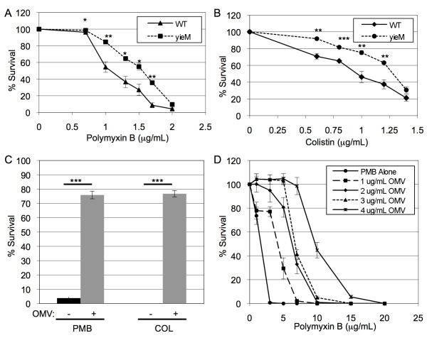
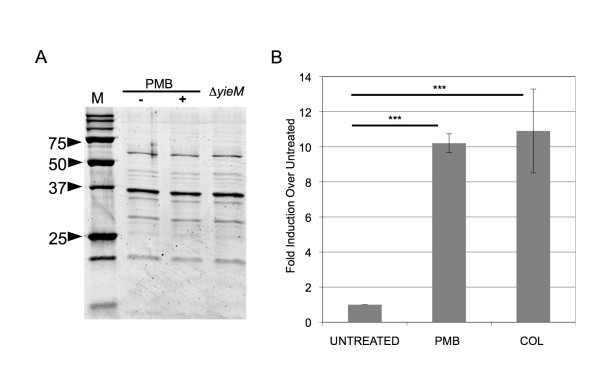
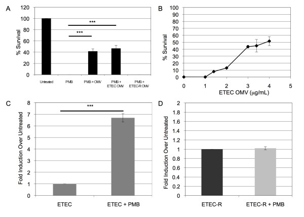
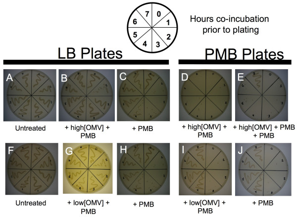
Results: We found that a hyper-vesiculating mutant of Escherichia coli survived treatment by antimicrobial peptides (AMPs) polymyxin B and colistin better than the wild-type. Supplementation of E. coli cultures with purified outer membrane vesicles provided substantial protection against AMPs, and AMPs significantly induced vesiculation. Vesicle-mediated protection and induction of vesiculation were also observed for a human pathogen, enterotoxigenic E. coli (ETEC), challenged with polymyxin B. When ETEC with was incubated with low concentrations of vesicles concomitant with polymyxin B treatment, bacterial survival increased immediately, and the culture gained resistance to polymyxin B. By contrast, high levels of vesicles also provided immediate protection but prevented acquisition of resistance. Co-incubation of T4 bacteriophage and OMVs showed fast, irreversible binding. The efficiency of T4 infection was significantly reduced by the formation of complexes with the OMVs.

Conclusions: These data reveal a role for OMVs in contributing to innate bacterial defense by adsorption of antimicrobial peptides and bacteriophage. Given the increase in vesiculation in response to the antimicrobial peptides, and loss in efficiency of infection with the T4-OMV complex, we conclude that OMV production may be an important factor in neutralizing environmental agents that target the outer membrane of Gram-negative bacteria.


Figures

Figure 1
Figure 1
OMV-mediated protection to AMPs. Relative survival of WT (solid line) and ΔyieM (dashed line) E.coli after 2 h treatment with the indicated concentrations of polymyxin B (A) and colistin (B). (C) Cultures of mid-log phase WT E. coli were simultaneously treated with the indicated antibiotic (polymyxin B (PMB) 1.5 μg/mL and colistin (COL) 1.0 μg/mL) and either no OMVs (black bars) or with OMVs purified from WT E.coli (4 μg/mL) (grey bars). (D) To titrate OMV-mediated protection, indicated concentrations of WT OMVs were co-incubated in media for 2 h with indicated concentrations of polymyxin B and the media cleared of OMVs by centrifugation. Polymyxin B activity remaining in the media was assessed by adding the pretreated media to a mid log-phase culture of WT E. coli, incubating for 2 h, and plating for CFU. Relative growth (% Survival) was determined by dividing the CFU/mL obtained from antibiotic-treated cultures by the CFU/mL from cultures without antibiotic. (n = 9 for all experiments).
Figure 2
Figure 2
OMV production is substantially induced by AMPs. (A) OMVs from 0.75 μg/mL polymyxin B-treated (+) and untreated (-) WT cultures were purified, separated by SDS-PAGE, and stained using SYPRO Ruby Red. OMVs from strain ΔyieM are also shown for comparison. No significant differences in protein content could be identified across all samples. Molecular weight standards are indicated in kDa (M). (B) OMVs in the cell-free culture supernatant of antibiotic-treated WT cultures (0.75 μg/mL polymyxin B, PMB; or 0.5 μg/mL colistin, COL) were quantitated by measuring outer membrane protein and compared with the quantity of OMVs produced by untreated cultures (Untreated). Production was normalized to CFU/mL of each culture at the time of OMV preparation, and relative fold-differences are shown. (n = 9 for all experiments).
Figure 3
Figure 3
ETEC, not ETEC-R, OMVs are protective and induced by polymyxin B. (A) A mid-log culture of ETEC was treated with polymyxin B (4 μg/mL, final concentration) simultaneous with no addition (PMB), 10 μg of WT (K12) OMVs (PMB + OMV), 10 μg of ETEC-derived OMVs (PMB + ETEC OMV), or 10 μg ETEC-R-derived OMVs (PMB + R-ETEC-OMV). Relative growth (% Survival) was determined compared to cultures without antibiotic (Untreated). (n = 9) (B) To titrate OMV-mediated protection for ETEC, ETEC OMVs (final concentrations indicated) were added simultaneously with polymyxin B (5 μg/mL, final concentration) to a mid-log phase ETEC culture and co-incubated 2 h at 37°C. Relative growth (% Survival) was determined compared to cultures without antibiotic. (n = 6) OMV yield was quantitated for mid-log phase cultures of ETEC (C) or ETEC-R (D) treated for 14 h with 3 μg/ml polymyxin B. (n = 6 for both C and D) OMV production was normalized to the CFU/mL of each culture at the time of vesicle harvest, and relative fold-differences compared to untreated cultures are shown.
Figure 4
Figure 4
Acquisition of ETEC resistance to polymyxin B is reduced by co-incubation with high concentrations of OMVs. At hourly time-points for 0-7 h of co-incubation, equivalent volumes of the samples described below were streaked on each plate in a pattern indicated by the template diagram. Top row: ETEC co-incubated with (A) nothing, (B, D) a high concentration of ETEC OMV (2 μg/mL) and polymyxin B (3.5 μg/ml), or (C) polymyxin B alone (3.5 μg/mL). Samples were streaked either on LB agar (A-C), or LB containing 5 μg/ml polymyxin B (D-E). (E) ETEC co-incubated with ETEC OMV (3 μg/mL) and polymyxin B (3.5 μg/mL) for 5 h, then an additional 5 μg/mL polymyxin B was added, and plated on LB containing 5 μg/mL polymyxin B. Resistance was seen by hour 7 without decreasing cell population significantly. Bottom row: ETEC co-incubated with (F) nothing, or (G, I) 1.4 μg/mL ETEC OMV and 3.5 μg/ml polymyxin B, and (H, J) polymyxin B alone (3.5 μg/mL), streaked on LB (F-H) or LB containing 5 μg/mL polymyxin B (I-J). (n = 9 for all experiments).
Figure 5
Figure 5
T4 phage bind OMVs, reducing their capacity to infect E. coli. (A) 106 T4 phage were co-incubated with 1 μg purified WT OMVs (106 T4+OMV) for 2 h. As controls, 106 T4, 1 μg of purified WT OMVs, and 105 T4 were also incubated under the same conditions for 2 h. For the 5 min panel, samples were mixed with MK496 cells and allowed to incubate for 5 min, PFU were then determined and compared to the PFU produced by the 106 T4 sample (% PFU Remaining). For the 60 min panel, the phage and WT OMV preparations were incubated for 1 h with mid log-phase MK496 cells, PFU were determined, and compared to the PFU produced by the 106 T4 sample (% PFU Remaining). (n = 9) (B) 106 T4 phage were mixed with 1 μg purified WT OMVs, then immediately ("0" min), and at 5 min intervals thereafter, samples were taken and chloroform was added to disrupt the OMVs and allow reversibly bound phage to be released. The T4 activity in each sample was determined by PFU titration and compared to the PFU produced by 106 T4 (% PFU Remaining). (n = 6) (C) Negative stain electron micrograph of the T4-OMV complex (size bar = 50 nm).

References

    1. McDermott PF, Walker RD, White DG. Antimicrobials: modes of action and mechanisms of resistance. Int J Toxicol. 2003;22(2):135–143. doi: 10.1080/10915810305089. - DOI - PubMed
    1. Kulp A, Kuehn MJ. Biological functions and biogenesis of secreted bacterial outer membrane vesicles. Annu Rev Microbiol. 2010;64:163–184. doi: 10.1146/annurev.micro.091208.073413. - DOI - PMC - PubMed
    1. Ellis TN, Kuehn MJ. Virulence and immunomodulatory roles of bacterial outer membrane vesicles. Microbiol Mol Biol Rev. 2010;74(1):81–94. doi: 10.1128/MMBR.00031-09. - DOI - PMC - PubMed
    1. Kobayashi H, Uematsu K, Hirayama H, Horikoshi K. Novel toluene elimination system in a toluene-tolerant microorganism. J Bacteriol. 2000;182(22):6451–6455. doi: 10.1128/JB.182.22.6451-6455.2000. - DOI - PMC - PubMed
    1. Yonezawa H, Osaki T, Kurata S, Fukuda M, Kawakami H, Ochiai K, Hanawa T, Kamiya S. Outer membrane vesicles of Helicobacter pylori TK1402 are involved in biofilm formation. BMC Microbiol. 2009;9:197. doi: 10.1186/1471-2180-9-197. - DOI - PMC - PubMed

Publication types

MeSH terms

LinkOut - more resources