. 2011 Dec 1;4(1):26.
doi: 10.1186/1755-1536-4-26.

Animal models for investigating chronic pancreatitis

Affiliations

Animal models for investigating chronic pancreatitis

Alexander A Aghdassi et al. Fibrogenesis Tissue Repair. .

Abstract

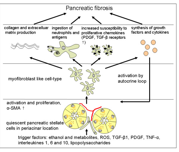
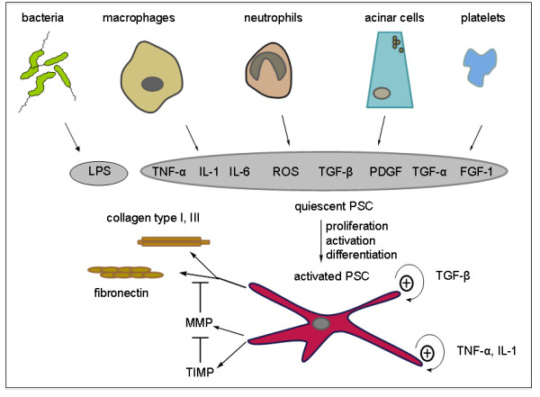
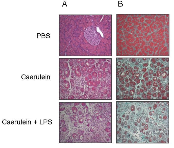
Chronic pancreatitis is defined as a continuous or recurrent inflammatory disease of the pancreas characterized by progressive and irreversible morphological changes. It typically causes pain and permanent impairment of pancreatic function. In chronic pancreatitis areas of focal necrosis are followed by perilobular and intralobular fibrosis of the parenchyma, by stone formation in the pancreatic duct, calcifications in the parenchyma as well as the formation of pseudocysts. Late in the course of the disease a progressive loss of endocrine and exocrine function occurs. Despite advances in understanding the pathogenesis no causal treatment for chronic pancreatitis is presently available. Thus, there is a need for well characterized animal models for further investigations that allow translation to the human situation. This review summarizes existing experimental models and distinguishes them according to the type of pathological stimulus used for induction of pancreatitis. There is a special focus on pancreatic duct ligation, repetitive overstimulation with caerulein and chronic alcohol feeding. Secondly, attention is drawn to genetic models that have recently been generated and which mimic features of chronic pancreatitis in man. Each technique will be supplemented with data on the pathophysiological background of the model and their limitations will be discussed.


Figures

Figure 1
Figure 1
Role of pancreatic stellate cells (PSCs) in fibrosis. Activation of PSCs from a quiescent into a myofibroblast-like type and proliferation occurs via several triggers and is perpetuated in an autocrine loop. Development of fibrosis is a complex process that requires interaction of collagen and extracellular matrix production together with chemokine synthesis and phagocytosis.
Figure 2
Figure 2
Multiple activators for pancreatic stellate cells. Adjacent acinar cells as well as neutrophils, macrophages and platelets stimulate pancreatic stellate cells (PSCs) in a paracrine manner involving growth factors, cytokines and reactive oxygen species (ROS). Bacterial endotoxin lipopolysaccharide (LPS) acts on PSCs. Once activated there is autocrine stimulation perpetuated by transforming growth factor β (TGFβ), tumor necrosis factor α (TNFα) and interleukin 6 (IL-6). They further express extracellular matrix components such as collagens I, III and fibronectin, and matrix metalloproteinases as well as their inhibitors.
Figure 3
Figure 3
Lipopolysaccharide (LPS)-dependent and nuclear factor (NF)κB-mediated activation of proinflammatory cytokines. LPS binds to LPS-binding protein (LBP) and forms a complex with the surface protein CD14 that further interacts with MD-2 associated Toll-like receptor 4 (TLR4). Myeloid differentiation primary response gene 88 (MyD88) is recruited to TLR4 by its Toll-interleukin 1 receptor (IL1R) (TIR) domain and further activates serine/threonine kinase interleukin 1 receptor-associated kinase (IRAK) by its death domain (DD). After recruitment of tumor necrosis factor receptor associated factor 6 (TRAF-6), another adapter protein, mitogen-activated protein kinase kinase kinase (MAPKKK) transforming growth factor β activated kinase (TAK1) is activated that phosphorylates the inhibitor of κB kinase (IKK) complex. Activated IKK removes inhibitory IκB from the IκB-NFκB complex so that NFκB can enter the nucleus and where it stimulates the expression of proinflammatory chemokines. In a second pathway, which is not listed here, TAK1 is able to activate MAP kinases p38 and c-Jun N-terminal kinase (JNK).
Figure 4
Figure 4
Histological changes of pancreatic tissue architecture after repetitive caerulein and combined caerulein-lipopolysaccharide (LPS) injections. (A) Hematoxylin and eosin staining indicates destruction of pancreatic acinar cells and development of tubular complexes, a sign of chronic pancreatitis. (B) Masson-Goldner trichrome stain denotes fibrosis; connective tissue is stained green. Caerulein and LPS increase pancreatic fibrosis compared to caerulein alone. Phosphate-buffered saline (PBS)-treated mice did not develop chronic pancreatitis and were used as negative controls.

References

    1. Ammann RW, Akovbiantz A, Largiader F, Schueler G. Course and outcome of chronic pancreatitis. Longitudinal study of a mixed medical-surgical series of 245 patients. Gastroenterology. 1984;86:820–828. - PubMed
    1. Etemad B, Whitcomb DC. Chronic pancreatitis: diagnosis, classification, and new genetic developments. Gastroenterology. 2001;120:682–707. doi: 10.1053/gast.2001.22586. - DOI - PubMed
    1. Sarner M, Cotton PB. Classification of pancreatitis. Gut. 1984;25:756–759. doi: 10.1136/gut.25.7.756. - DOI - PMC - PubMed
    1. Chari ST, Singer MV. The problem of classification and staging of chronic pancreatitis. Proposals based on current knowledge of its natural history. Scand J Gastroenterol. 1994;29:949–960. doi: 10.3109/00365529409094869. - DOI - PubMed
    1. Andersen BN, Pedersen NT, Scheel J, Worning H. Incidence of alcoholic chronic pancreatitis in Copenhagen. Scand J Gastroenterol. 1982;17:247–252. doi: 10.3109/00365528209182047. - DOI - PubMed

LinkOut - more resources