

High-level mucosal and systemic immune responses induced by oral administration with Lactobacillus-expressed porcine epidemic diarrhea virus (PEDV) S1 region combined with Lactobacillus-expressed N protein
- PMID: 22134641
- PMCID: PMC7080084
- DOI: 10.1007/s00253-011-3734-0
High-level mucosal and systemic immune responses induced by oral administration with Lactobacillus-expressed porcine epidemic diarrhea virus (PEDV) S1 region combined with Lactobacillus-expressed N protein
Abstract
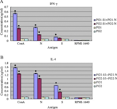
To develop effective mucosal vaccine formulation against porcine epidemic diarrhea virus (PEDV) infection, the DNA fragments encoding spike protein immunodominant region S1 and nucleocapsid N of PEDV were inserted into pPG1 (surface-displayed) or pPG2 (secretory) plasmids followed by electrotransformation into Lactobacillus casei (Lc) to yield four recombinant strains: PG1-S1, PG2-S1, PG1-N, and PG2-N. After intragastric administration, it was observed that live Lc-expressing S1 protein combined with Lc-expressing N protein could elicit much more potent mucosal and systemic immune responses than the former alone (P < 0.001), however slightly inferior to the latter alone (P > 0.05). Furthermore, the surface-displayed mixture (PG1-S1+ PG1-N) revealed stronger immunogenicity than the secretory mixture (PG2-S1+ PG2-N) as well as PEDV-neutralizing potency in vitro (P < 0.001). On 49th day after the last immunization, splenocytes were prepared from mice immunized with surface-displayed mixture, secretory mixture and negative control to be stimulated by purified N and S protein, respectively. The results of ELISA analysis showed that N protein was capable of inducing a higher level of IL-4 (P < 0.001) and IFN-γ (P < 0.001) than S1 protein in the immunized mice. Taken together, Lc-expressed N protein as molecular adjuvant or immunoenhancer was able to effectively facilitate the induction of mucosal and systemic immune responses by Lc-expressing S1 region.
Figures


References
-
- BaoXian L, GuangPeng M, JunWei G, YiJing L. Porcine aminopeptidase N is a functional receptor for the PEDV coronavirus. Chin J Virol. 2009;25(3):220–225. - PubMed
-
- Butler JE. Immunoglobulins and immunocytes in animal milks. In: Orga P, Mestecky J, Lamm M, Strober W, Bienenstock J, McGhee JR, editors. Mucosal immunology. 2. New York: Academic; 1999. pp. 1531–1534.
-
- Claassen E, Van Winsen R, Posno M, Boersma WJ. New and safe "oral" live vaccines based on Lactobacillus. Adv Exp Med Biol. 1995;371B:1553–1558. - PubMed
-
- Collins LV, Schodel F. Live bacterial vector. In: Verschueren PC, editor. Pastorel PP, Blancou Vannier J. New York: Veterinary vaccinology. Elsevier Science; 1997. pp. 293–308.
Publication types
MeSH terms
Substances
LinkOut - more resources
Full Text Sources
Other Literature Sources
