Review
. 2012 Apr;16(2):214-20.
doi: 10.1007/s10157-011-0568-0. Epub 2011 Dec 2.

Involvement of glomerular renin-angiotensin system (RAS) activation in the development and progression of glomerular injury

Affiliations
Review

Involvement of glomerular renin-angiotensin system (RAS) activation in the development and progression of glomerular injury

Shoji Kagami. Clin Exp Nephrol. 2012 Apr.

Abstract

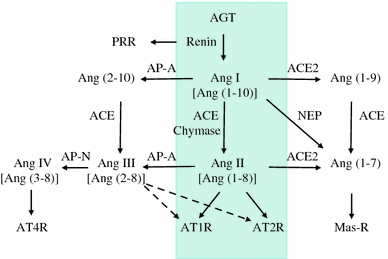
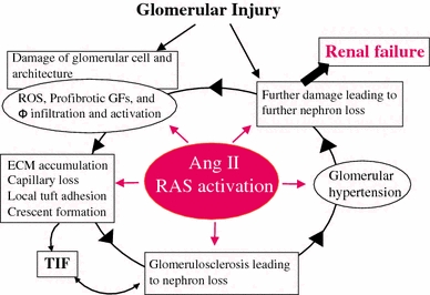
Recently, there has been a paradigm shift away from an emphasis on the role of the endocrine (circulating) renin-angiotensin system (RAS) in the regulation of the sodium and extracellular fluid balance, blood pressure, and the pathophysiology of hypertensive organ damage toward a focus on the role of tissue RAS found in many organs, including kidney. A tissue RAS implies that RAS components necessary for the production of angiotensin II (Ang II) reside within the tissue and its production is regulated within the tissue, independent of the circulating RAS. Locally produced Ang II plays a role in many physiological and pathophysiological processes such as hypertension, inflammation, oxidative stress, and tissue fibrosis. Both glomerular and tubular compartments of the kidney have the characteristics of a tissue RAS. The purpose of this article is to review the recent advances in tissue RAS research with a particular focus on the role of the glomerular RAS in the progression of renal disease.


Figures

Fig. 1
Fig. 1
Overview of the renin−angiotensin system (RAS). The schematic shows the circulating RAS (inside the four-sided line) as well as newly recognized enzymatic pathways that lead to the formation and metabolism of products derived from angiotensinogen (AGT). PRR prorenin/renin receptor, ACE angiotensin-converting enzyme, ACE2 angiotensin-converting enzyme 2, AP-A aminopeptidase A, AP-N aminopeptidase N, NEP neprilysin, Ang I angiotensin I, Ang II angiotensin II, AT1R angiotensin II type I receptor, AT2R angiotensin II type 2 receptor, AT4R angiotensin II type 4 receptor. Modified from Refs. [9, 10]
Fig. 2
Fig. 2
The central role of angiotensin II (RAS activation) in progressive glomerular injury. ROS reactive oxygen species, GFs growth factors, Φ macrophage, TIF tubulo-interstitial fibrosis; ECM, extracellular matrix. Modified from Refs. [18, 20]
Fig. 3
Fig. 3
Protein expression of angiotensinogen (AGT) in isolated human glomeruli and immunohistochemical staining of AGT in patients with minor glomerular abnormalities (MGA) or IgA nephropathy (IgAN). a Western blot analysis was performed using samples of isolated human glomeruli (lane 1) and purified human AGT (lane 2), respectively. Anti-human AGT antibody reacted with a 61 kDa band in each sample. b In patients with MGA, AGT was strongly expressed in proximal tubules and weakly detected in glomerular endothelial cells. c In patients with IgAN, AGT expression was strongly induced by glomerular endothelial cells and mesangial cells. Modified from Ref. [30]
Fig. 4
Fig. 4
Effects of the ARB candesartan in anti-GBM antibody-induced nephritic rats. Nephritic rats were treated with or without candesartan, sacrificed on day 28, and then subjected to an immunohistochemical examination. Light microscopic examination showed that severe crescentic nephritis had developed by day 28 (b) but was significantly attenuated by treatment with ARB (c). PBS-injected rats were used as normal control rats (a, d, g). Immunostaining revealed that nephritic rats showed diffuse and strong glomerular Ang II staining (e), while ARB treated-nephritic rats showed segmental accentuated staining of Ang II (f). Control rats showed weak positive Ang II staining (d). Strong superoxide production (DHE dye) was detected in nephritic rats (h) compared with control rats (g), but was significantly attenuated in ARB treated-nephritic rats (i). Modified from Ref. [39]
Fig. 5
Fig. 5
Biochemical analysis of nephritic rats on day 28 with or without treatment with ARB. Samples from isolated glomeruli from either control rats, day 28 nephritic rats or ARB-treated day 28 nephritic rats were subjected to Western blot analysis using anti-AGT antibody (a), Ang II measurement using ELISA (b), TGF-β measurement using ELISA (c) and Western blot analysis using anti-Nox2 antibody (d). Control control rats, GN nephritic rats without ARB treatment, GN + ARB nephritic rats with ARB treatment. # p < 0.01 versus control; § p < 0.05 versus GN; *p < 0.01 versus GN. Modified from Ref. [39]

Similar articles

Cited by

References

    1. Tigerstedt R, Bergman PG. Niere und Kreislauf. Skand Arch Physiol. 1898;8:223–271.
    1. Bader M. Tissue renin-angiotensin-aldosterone systems: targets for pharmacological therapy. Annu Rev Pharmacol Toxicol. 2010;50:439–465. doi: 10.1146/annurev.pharmtox.010909.105610. - DOI - PubMed
    1. Dzau VJ. Tissue angiotensin and pathobiology of vascular disease: a unifying hypothesis. Hypertension. 2001;37:1047–1052. - PubMed
    1. Bader M, Ganten D. Update on tissue renin-angiotensin systems. J Mol Med (Berl) 2008;86:615–621. doi: 10.1007/s00109-008-0336-0. - DOI - PubMed
    1. Suzuki Y, Ruiz-Ortega M, Lorenzo O, Ruperez M, Esteban V, Egido J. Inflammation and angiotensin II. Int J Biochem Cell Biol. 2003;35:881–900. doi: 10.1016/S1357-2725(02)00271-6. - DOI - PubMed

Publication types