. 2012 Feb 17;287(8):5211-24.
doi: 10.1074/jbc.M111.290080. Epub 2011 Dec 1.

Dentin phosphoprotein (DPP) activates integrin-mediated anchorage-dependent signals in undifferentiated mesenchymal cells

Affiliations

Dentin phosphoprotein (DPP) activates integrin-mediated anchorage-dependent signals in undifferentiated mesenchymal cells

Asha Eapen et al. J Biol Chem. .

Abstract

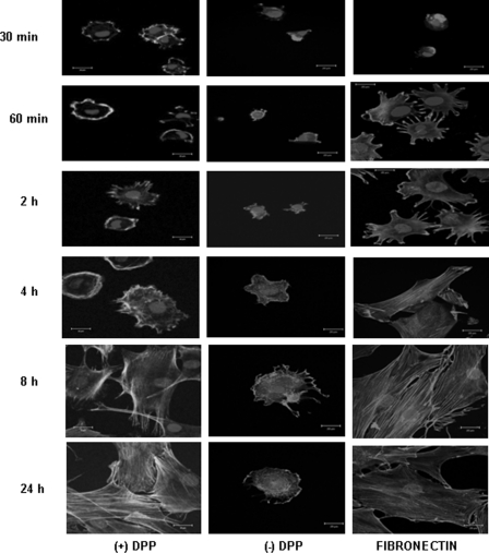
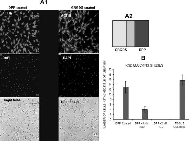
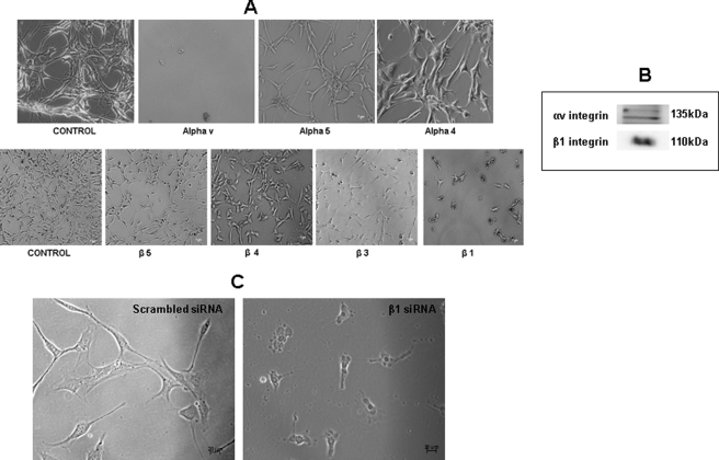
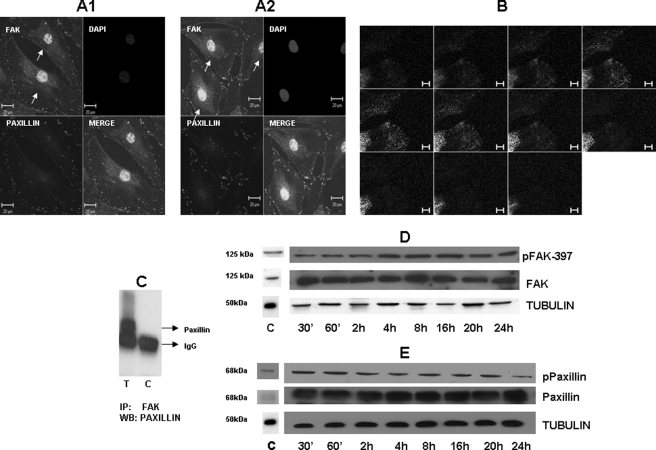
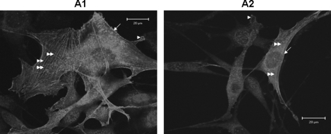
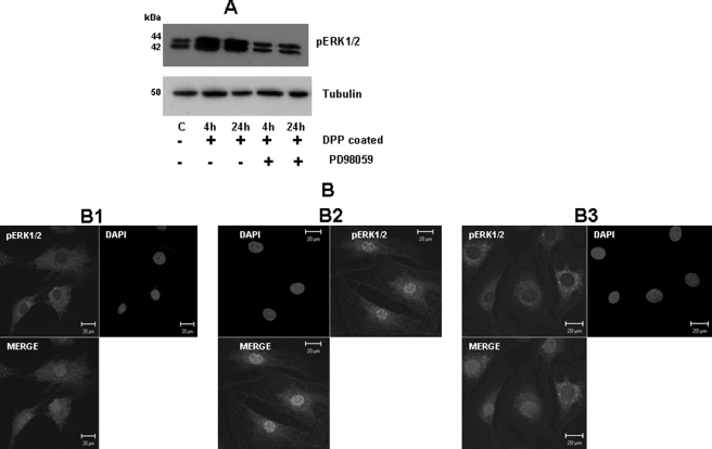
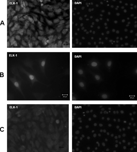
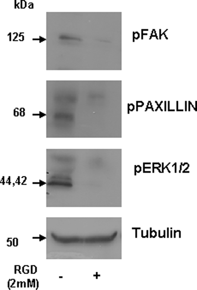
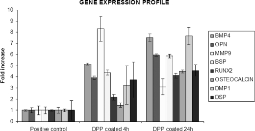
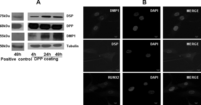
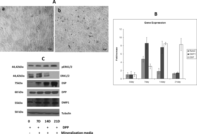
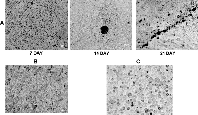
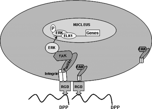
Dentin phosphoprotein (DPP), a major noncollagenous protein of the dentin matrix, is a highly acidic protein that binds Ca(2+) avidly and is thus linked to matrix mineralization. Here, we demonstrate that the RGD domain in DPP can bind to integrins on the cell surface of undifferentiated mesenchymal stem cells and pulp cells. This coupling generates intracellular signals that are channeled along cytoskeletal filaments and activate the non-receptor tyrosine kinase focal adhesion kinase, which plays a key role in signaling at sites of cellular adhesion. The putative focal adhesion kinase autophosphorylation site Tyr(397) is phosphorylated during focal adhesion assembly induced by DPP on the substrate. We further demonstrate that these intracellular signals propagate through the cytoplasm and activate anchorage-dependent ERK signaling. Activated ERK translocates to the nucleus and phosphorylates the transcription factor ELK-1, which in turn coordinates the expression of downstream target genes such as DMP1 and dentin sialoprotein (DSP). These studies suggest a novel paradigm demonstrating that extracellular DPP can induce intracellular signaling that can be propagated to the nucleus and thus alter gene activities.


Figures

FIGURE 1.
FIGURE 1.
DPP on substrate increases C3H10T1/2 cell spreading and adhesion. C3H10T1/2 cells were seeded on DPP- or non-DPP-coated (()DPP) coverglass for 30 min to 24 h. The cells were fixed and stained for actin. Cells seeded on fibronectin served as a positive control. Scale bars, 20 μm.
FIGURE 2.
FIGURE 2.
RGD domain in DPP facilitates cell attachment. Gold substrate was stamped with DPP on one side and an RGD peptide on the other side. C3H10T1/2 cells were seeded on the substrate for 4 h, fixed, and stained with actin (A1). The stamping pattern is schematically represented in A2. C3H10T1/2 cells were incubated with a 1 or 2 mm concentration of RGD blocking peptide for 2 h. Cells were then allowed to attach on a DPP-coated coverglass and manually counted with a hemocytometer. The bar graph represents the number of attached cells (B). Scale bar, 50 μm. Error bars represent mean ± standard deviation (n = 3).
FIGURE 3.
FIGURE 3.
Identification of the cell surface α and β integrins specific for DPP ligand. Receptor-cell binding experiments were performed by incubating C3H10T1/2 cells with either αv, α4, α5, β5, β4, β3, or β1 antibody for 2 h, respectively. The combinations of cells with the integrin antibodies were seeded on DPP-coated plates and incubated for 24 h. Light microscopic images (Zeiss) of the cells on DPP-coated plates were imaged (A). Note the specificity of the αv and β1 antibodies. Specificity was confirmed by isolating membrane proteins, and Western blotting was performed using αv and β1 integrin antibodies (B). β1 integrin siRNA transfection was performed in C3H10T1/2 cells grown on DPP-coated plates. 0.5 μg of the siRNA was used for transfection. Transfection performed with scrambled siRNA was used as the control (C).
FIGURE 4.
FIGURE 4.
DPP triggers organization of focal adhesion complexes. C3H10T1/2 cells were seeded on DPP-coated plates for 16 (A1) and 24 h (A2). Immunohistochemical analysis was performed with paxillin (red) and FAK (green) antibodies. Confocal microscopic images are presented in A. Immunostaining indicates the successful spreading and formation of focal adhesions. FAK is localized at focal adhesions and the nucleus (single arrows), whereas paxillin is localized predominantly at focal adhesions. Scale bars, 20 μm. Z stack analysis was performed to determine the colocalization of FAK with paxillin at the 24-h time point (B). DPP induces in vivo association of FAK and paxillin (C). “T” represents 50 μg of total protein and FAK antibody mixed with protein A beads and incubated overnight. The beads were then washed with PBS, and SDS-PAGE loading dye was added to the beads and boiled. Immunoblot was performed with paxillin antibody. “C” represents immunoprecipitation (IP) performed in the absence of FAK antibody. C3H10T1/2 cells were seeded on DPP-coated plates, total proteins were isolated at various time points as indicated, and Western blotting (WB) was performed to determine activation of FAK (D) and paxillin (E). Total proteins isolated from C3H10T1/2 cells grown in the absence of DPP were used as a control (C). pPaxillin, phosphopaxillin; pFAK, phospho-FAK; ′, minutes.
FIGURE 5.
FIGURE 5.
Cells on DPP substrate promote actin polymerization. C3H10T1/2 cells were transfected with an mRFPruby-actin plasmid using FuGENE HD. The cells were then trypsinized and seeded on a DPP-coated coverglass. Confocal microscopic images were taken at 8 (A1) and 24 h (A2). Note the incorporation of the mRFPruby-actin into subcompartments of the actin cytoskeleton such as lamellipodium (single arrows), filopodia (single arrowheads), and stress fibers (double arrowheads). Scale bars, 20 μm.
FIGURE 6.
FIGURE 6.
Activation of ERK1/2 MAPK pathway by adherent cells on DPP substrate. C3H10T1/2 cells were seeded on DPP-coated plates and treated with or without PD98059 for 4 and 24 h. Total proteins were isolated, and Western blotting was performed using phospho-ERK1/2 (pERK1/2) and tubulin antibodies. Cells in suspension were used as a control (A). Confocal imaging was performed on adherent cells at 24 h. No nuclear localization was observed in cells seeded on tissue culture coverglass (B1). Nuclear localization of phospho-ERK1/2 was seen in cells plated on DPP-coated coverglass (B2), and this was inhibited after PD98059 treatment for 24 h (B3). C, control. Scale bars, 20 μm.
FIGURE 7.
FIGURE 7.
Cells adherent on DPP stimulate nuclear accumulation of ELK-1 transcription factor. A confocal image of ELK-1 immunostaining performed on C3H10T1/2 cells seeded on DPP-coated coverglass is shown in A. A transient transfection of a FLAG construct of ELK-1 was performed using Superfect reagent on C3H10T1/2 cells seeded on DPP-coated plates. Localization of ELK-1 was determined by immunofluorescence with an ELK-1 antibody and TRITC-conjugated anti-rabbit secondary antibody. Nuclear localization of ELK-1 is seen in the DPP-treated plates (B). Cells seeded on a tissue culture plate were used as a control (C). Scale bars, 20 μm.
FIGURE 8.
FIGURE 8.
Blocking RGD abrogates activation of FAK, paxillin, and ERK1/2. C3H10T1/2 cells were incubated with 2 mm RGD peptide at room temperature for 2 h. The cells were then seeded on DPP-coated plates. Total protein was isolated after 24 h, and immunoblotting was performed with anti-phospho-ERK1/2 (pERK-1/2), phospho-FAK (pFAK), phosphopaxillin (pPaxillin), and tubulin antibodies. Cells seeded on a DPP-coated plate were used as a control.
FIGURE 9.
FIGURE 9.
DPP induces odontogenic differentiation in C3H10T1/2 cells. C3H10T1/2 cells were seeded on DPP-coated plates for 4 and 24 h. Total RNA was isolated, subjected to real time PCR, and analyzed for the expression of RUNX2, DSP, DMP1, BMP4, BSP, OPN, MMP9, and osteocalcin (OCN). Error bars represent mean ± standard deviation (n = 3). GAPDH was used as the housekeeping gene.
FIGURE 10.
FIGURE 10.
DPP triggers nuclear localization and protein synthesis of odontogenic markers of differentiation. Total protein was isolated from cells seeded on DPP-coated plates at 4, 24, and 48 h, and immunoblotting was performed with DMP1, DSP, DPP, and tubulin antibodies (A). Cells seeded on a tissue culture plate for 48 h were used as a control. A confocal image of C3H10T1/2 cells seeded on DPP-coated coverglass for 24 h and immunostained using antibodies for DSP, DMP1, and RUNX2 is shown (B). Nuclear localization of DSP, DMP1, and RUNX2 was seen in cells plated on DPP-coated coverglass. +ve, positive; pERK1/2, phospho-ERK1/2.
FIGURE 11.
FIGURE 11.
DPP plays important role in terminal differentiation of odontoblast. C3H10T1/2 cells seeded on DPP-coated plates were grown in mineralization medium for 7, 14, and 21 days. Light microscopic images of the cells in the presence and absence of mineralization medium at 21 days were taken (A). Changes in the morphology of the cells were observed in the absence (A, panel a) and presence (A, panel b) of mineralization medium. Scale bars, 20 μm. RNA was extracted using TRIzol at 7, 14, and 21 days, and real time PCR was performed for RUNX2, DSP, and DMP1 (B). Total proteins were isolated from C3H10T1/2 cells seeded on DPP-coated plates and grown under mineralization conditions for 7, 14, and 21 days (C). Immunoblotting was performed with DSP, DMP1, DPP, and phospho-ERK1/2 (pERK1/2). Total proteins isolated from cells grown on DPP-coated plates for 21 days in the absence of mineralization medium were used as control. Error bars represent mean ± standard deviation.
FIGURE 12.
FIGURE 12.
DPP triggers mineralized nodule formation in C3H10T1/2 cells. von Kossa staining was performed on C3H10T1/2 cells seeded on DPP-coated plates grown in mineralization medium for 7, 14, and 21 days (A). The cells were fixed in formalin, washed twice with distilled water, and then stained with 1% silver nitrate solution. von Kossa staining demonstrated a high intensity of dark staining pattern at 14 and 21 days of culture. Cells grown on DPP-coated plates without mineralization medium (B) and cells on a tissue culture plate with mineralization medium (C) were used as controls. Scale bars, 20 μm.
FIGURE 13.
FIGURE 13.
DPP induces matrix mineralization in C3H10T1/2 cells. C3H10T1/2 cells seeded on DPP-coated plates were grown in the presence of mineralization medium for 7, 14, and 21 days (A). Matrix mineralization is indicated by the dark areas as a result of calcium staining with Alizarin Red. As controls, cells were grown in mineralization conditions on tissue culture plates for 7, 14, and 21 days (B). Scale bars, 20 μm.
FIGURE 14.
FIGURE 14.
Hypothetical model. The hypothetical model depicts the RGD-mediated integrin anchorage of cells on DPP and the subsequent activation of focal adhesion complexes and downstream gene transcription.

References

    1. Feng J. Q., Luan X., Wallace J., Jing D., Ohshima T., Kulkarni A. B., D'Souza R. N., Kozak C. A., MacDougall M. (1998) Genomic organization, chromosomal mapping, and promoter analysis of the mouse dentin sialophosphoprotein (Dspp) gene, which codes for both dentin sialoprotein and dentin phosphoprotein. J. Biol. Chem. 273, 9457–9464 - PubMed
    1. Yamakoshi Y., Lu Y., Hu J. C., Kim J. W., Iwata T., Kobayashi K., Nagano T., Yamakoshi F., Hu Y., Fukae M., Simmer J. P. (2008) Porcine dentin sialophosphoprotein: length polymorphisms, glycosylation, phosphorylation, and stability. J. Biol. Chem. 283, 14835–14844 - PMC - PubMed
    1. George A., Srinivasan R., Thotakura S. R., Liu K., Veis A. (1999) Rat dentin matrix protein 3 is a compound protein of rat dentin sialoprotein and phosphophoryn. Connect. Tissue Res. 40, 49–57 - PubMed
    1. Yamakoshi Y., Hu J. C., Iwata T., Kobayashi K., Fukae M., Simmer J. P. (2006) Dentin sialophosphoprotein is processed by MMP-2 and MMP-20 in vitro and in vivo. J. Biol. Chem. 281, 38235–38243 - PubMed
    1. Yamakoshi Y., Hu J. C., Fukae M., Zhang H., Simmer J. P. (2005) Dentin glycoprotein: the protein in the middle of the dentin sialophosphoprotein chimera. J. Biol. Chem. 280, 17472–17479 - PubMed

Publication types

MeSH terms

LinkOut - more resources