

Molecular mechanism of active zone organization at vertebrate neuromuscular junctions
- PMID: 22135013
- PMCID: PMC3890249
- DOI: 10.1007/s12035-011-8216-y
Molecular mechanism of active zone organization at vertebrate neuromuscular junctions
Abstract
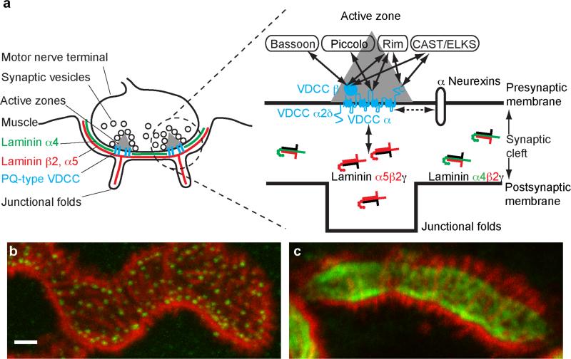
Organization of presynaptic active zones is essential for development, plasticity, and pathology of the nervous system. Recent studies indicate a trans-synaptic molecular mechanism that organizes the active zones by connecting the pre- and the postsynaptic specialization. The presynaptic component of this trans-synaptic mechanism is comprised of cytosolic active zone proteins bound to the cytosolic domains of voltage-dependent calcium channels (P/Q-, N-, and L-type) on the presynaptic membrane. The postsynaptic component of this mechanism is the synapse organizer (laminin β2) that is expressed by the postsynaptic cell and accumulates specifically on top of the postsynaptic specialization. The pre- and the postsynaptic components interact directly between the extracellular domains of calcium channels and laminin β2 to anchor the presynaptic protein complex in front of the postsynaptic specialization. Hence, the presynaptic calcium channel functions as a scaffolding protein for active zone organization and as an ion-conducting channel for synaptic transmission. In contrast to the requirement of calcium influx for synaptic transmission, the formation of the active zone does not require the calcium influx through the calcium channels. Importantly, the active zones of adult synapses are not stable structures and require maintenance for their integrity. Furthermore, aging or diseases of the central and peripheral nervous system impair the active zones. This review will focus on the molecular mechanisms that organize the presynaptic active zones and summarize recent findings at the neuromuscular junctions and other synapses.
Figures
Similar articles
-
Loss of β2-laminin alters calcium sensitivity and voltage-gated calcium channel maturation of neurotransmission at the neuromuscular junction.J Physiol. 2015 Jan 1;593(1):245-65. doi: 10.1113/jphysiol.2014.284133. Epub 2014 Dec 1. J Physiol. 2015. PMID: 25556799 Free PMC article.
-
Transsynaptic channelosomes: non-conducting roles of ion channels in synapse formation.Channels (Austin). 2011 Sep-Oct;5(5):432-9. doi: 10.4161/chan.5.5.16472. Epub 2011 Sep 1. Channels (Austin). 2011. PMID: 21654201 Free PMC article. Review.
-
Calcium channels link the muscle-derived synapse organizer laminin β2 to Bassoon and CAST/Erc2 to organize presynaptic active zones.J Neurosci. 2011 Jan 12;31(2):512-25. doi: 10.1523/JNEUROSCI.3771-10.2011. J Neurosci. 2011. PMID: 21228161 Free PMC article.
-
Measuring Presynaptic Calcium Influx at the Drosophila Larval Neuromuscular Junction.Cold Spring Harb Protoc. 2025 May 5;2025(5):pdb.prot108426. doi: 10.1101/pdb.prot108426. Cold Spring Harb Protoc. 2025. PMID: 38688542
-
Active zones of mammalian neuromuscular junctions: formation, density, and aging.Ann N Y Acad Sci. 2012 Dec;1274(1):24-32. doi: 10.1111/j.1749-6632.2012.06836.x. Ann N Y Acad Sci. 2012. PMID: 23252894 Free PMC article. Review.
Cited by
-
Skeletal muscle: A review of molecular structure and function, in health and disease.Wiley Interdiscip Rev Syst Biol Med. 2020 Jan;12(1):e1462. doi: 10.1002/wsbm.1462. Epub 2019 Aug 13. Wiley Interdiscip Rev Syst Biol Med. 2020. PMID: 31407867 Free PMC article. Review.
-
Presynaptic Active Zone Density during Development and Synaptic Plasticity.Front Mol Neurosci. 2012 Feb 15;5:12. doi: 10.3389/fnmol.2012.00012. eCollection 2012. Front Mol Neurosci. 2012. PMID: 22438837 Free PMC article.
-
Loss of β2-laminin alters calcium sensitivity and voltage-gated calcium channel maturation of neurotransmission at the neuromuscular junction.J Physiol. 2015 Jan 1;593(1):245-65. doi: 10.1113/jphysiol.2014.284133. Epub 2014 Dec 1. J Physiol. 2015. PMID: 25556799 Free PMC article.
-
Myasthenia Gravis: From the Viewpoint of Pathogenicity Focusing on Acetylcholine Receptor Clustering, Trans-Synaptic Homeostasis and Synaptic Stability.Front Mol Neurosci. 2020 May 28;13:86. doi: 10.3389/fnmol.2020.00086. eCollection 2020. Front Mol Neurosci. 2020. PMID: 32547365 Free PMC article.
-
Enhancement of Neuroglial Extracellular Matrix Formation and Physiological Activity of Dopaminergic Neural Cocultures by Macromolecular Crowding.Cells. 2022 Jul 6;11(14):2131. doi: 10.3390/cells11142131. Cells. 2022. PMID: 35883574 Free PMC article.
References
-
- Broadie KS, Richmond JE. Establishing and sculpting the synapse in Drosophila and C. elegans. Current Opinion in Neurobiology. 2002;12(5):491–498. - PubMed
-
- Collins CA, DiAntonio A. Synaptic development: insights from Drosophila. Current Opinion in Neurobiology. 2007;17(1):35–42. - PubMed
-
- Stryker E, Johnson KG. LAR, liprin alpha and the regulation of active zone morphogenesis. J Cell Sci. 2007;120(21):3723–3728. - PubMed
-
- Owald D, Sigrist SJ. Assembling the presynaptic active zone. Curr Opin Neurobiol. 2009;19(3):311–318. - PubMed
Publication types
MeSH terms
Substances
Grants and funding
LinkOut - more resources
Full Text Sources
