. 2012 Feb;53(2):311-20.
doi: 10.1194/jlr.M021865. Epub 2011 Nov 30.

Structural complex of sterol 14α-demethylase (CYP51) with 14α-methylenecyclopropyl-Delta7-24, 25-dihydrolanosterol

Affiliations

Structural complex of sterol 14α-demethylase (CYP51) with 14α-methylenecyclopropyl-Delta7-24, 25-dihydrolanosterol

Tatiana Y Hargrove et al. J Lipid Res. 2012 Feb.

Abstract

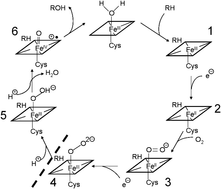
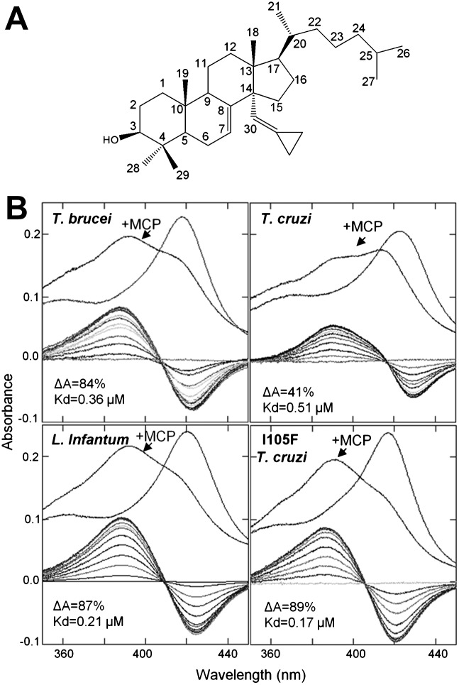
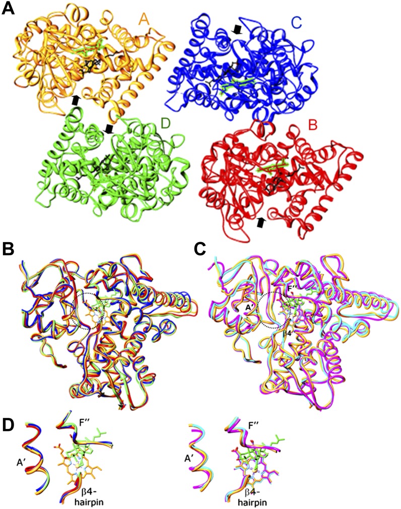
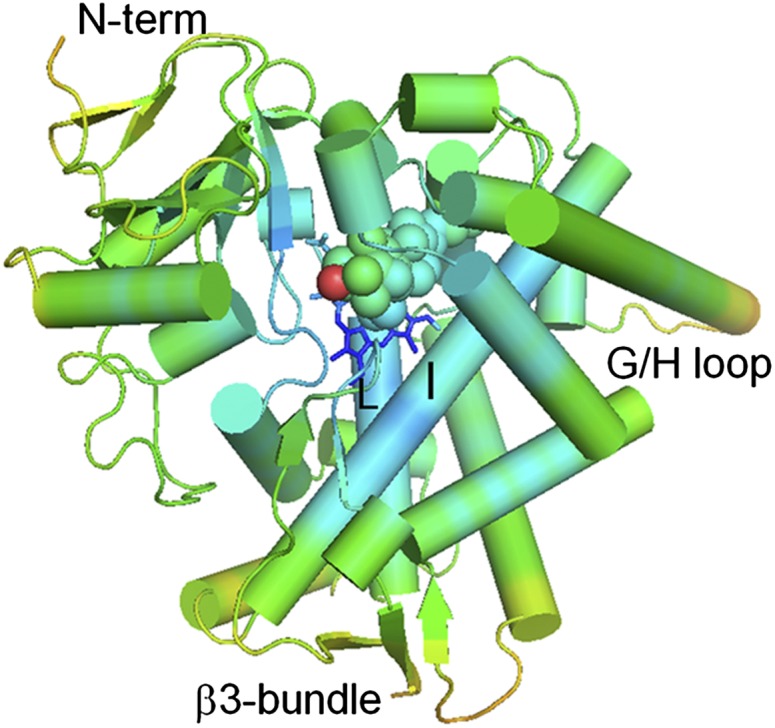
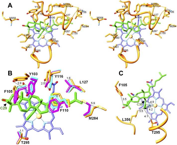
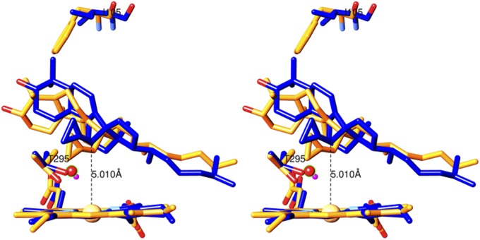
Sterol 14α-demethylase (CYP51) that catalyzes the removal of the 14α-methyl group from the sterol nucleus is an essential enzyme in sterol biosynthesis, a primary target for clinical and agricultural antifungal azoles and an emerging target for antitrypanosomal chemotherapy. Here, we present the crystal structure of Trypanosoma (T) brucei CYP51 in complex with the substrate analog 14α-methylenecyclopropyl-Δ7-24,25-dihydrolanosterol (MCP). This sterol binds tightly to all protozoan CYP51s and acts as a competitive inhibitor of F105-containing (plant-like) T. brucei and Leishmania (L) infantum orthologs, but it has a much stronger, mechanism-based inhibitory effect on I105-containing (animal/fungi-like) T. cruzi CYP51. Depicting substrate orientation in the conserved CYP51 binding cavity, the complex specifies the roles of the contact amino acid residues and sheds new light on CYP51 substrate specificity. It also provides an explanation for the effect of MCP on T. cruzi CYP51. Comparison with the ligand-free and azole-bound structures supports the notion of structural rigidity as the characteristic feature of the CYP51 substrate binding cavity, confirming the enzyme as an excellent candidate for structure-directed design of new drugs, including mechanism-based substrate analog inhibitors.


Figures

Fig. 1.
Fig. 1.
P450 catalytic cycle. Dashed line shows the step of the catalysis that is likely to be blocked in the MCP-bound T. brucei CYP51s due to disruption of the catalytic proton delivery (discussed in the text).
Fig. 2.
Fig. 2.
MCP. (A) Chemical structure (IUPAC carbon numbering). (B) Type 1 spectral responses in different CYP51s. Absolute (upper) and difference (lower) absorbance. P450 concentration, 1.8–2.0 μM; MCP concentration range, 0.25–5.0 μM. ΔA, spectral response amplitude (% of maximal low-to-high spin transition); Kd, apparent dissociation constant. Relative binding efficiencies (ΔA/Kd) are 1 for the wild-type T. cruzi (I105), 3 for T. brucei, 5 for L. infantum, and 6.5 for I105F T. cruzi CYP51s.
Fig. 3.
Fig. 3.
CYP51 in complex with MCP. The protein main chains are shown as cartoons; the heme (colored the same as the corresponding protein backbone) and MCP are seen as stick models. (A) Asymmetric unit. Four CYP51 protein chains are shown as gold, red, blue and, green ribbons (A, B, C, and D, respectively). Location of the substrate access channel entrance is marked with black arrow. The heme is framed in green. C-atoms of MCP are black. (B) Superimposition of the four MCP-CYP51 complexes from one asymmetric unit. Distal view. Average Cα rms deviation 0.638 Å. (C) MCP-bound (gold) versus ligand-free (cyan [3g1q]) versus VNI-bound (pink [3gw9]) CYP51. Substrate binding channel entrance is circled. MCP is colored in green; VNI is deleted for clarity. Binding of MCP causes fewer changes in the CYP51 backbone than binding of the azole inhibitor VNI. Average Cα rms deviation between the MCP-bound and VNI-bound CYP51 is 0.931 Å, whereas between the MCP-bound and ligand-free structures, it is only 0.653 Å. (D) Enlarged view of the secondary structural elements that form the access channel entrance. The substrate enters perpendicularly to the plane of the figure and then turns to acquire the catalytic position above the oxygen binding site. The colors and orientation of the CYP51molecules are the same as in B and C.
Fig. 4.
Fig. 4.
Heme-to-surface gradient of flexibility in the sterol-bound CYP51 molecule. The protein backbone is presented as a cartoon; heme is shown as sticks, MCP is seen as spheres. The C3 oxygen of MCP is red; all other atoms are colored by B-factors that increase from blue to orange. The coloring reveals the three most flexible regions in the complex: the GH-loop, the β3-bundle area, and the N-terminus, all located on the protein surface. The core of the molecule, including substrate binding cavity, displays the lowest flexibility.
Fig. 5.
Fig. 5.
MCP in the T. brucei CYP51 active site cavity. Stick model representation. The carbon atoms of MCP are green; the heme is blue. (A) Twenty CYP51 substrate contacting residues (stereoview). Except for M358, only side chains are shown. CYP51 family signature areas I (upper) and II (lower) are depicted as ribbons. (B) Altered side chain locations in the MCP-bound (gold) versus VNI-bound (pink) versus ligand-free CYP51 (cyan). Distances (Å) between the corresponding atoms in the MCP-bound and ligand-free CYP51 structures (dotted line) are shown. The C29 atom of MCP (the β-methyl carbon at C4) is marked with black arrow (C) The 14α-methylenecyclopropyl group of MCP contacts helix I (T295) and loop K/β1-4 (L356), which are presented as ribbons. The C30 atom of MCP (the 14α-methyl carbon) is marked with black arrow.
Fig. 6.
Fig. 6.
Superimposition of MCP in T. brucei (gold) and T. cruzi (blue) CYP51s (stereoview). MCP and the heme-coordinating water molecule (small pink sphere) from the MCP-bound and ligand-free T. brucei CYP51 structures, respectively, were modeled into the T. cruzi structure [3k1o] followed by energy minimization in Tripos (Sybyl). The MCP molecule in the T. cruzi CYP51 active site appears to be slightly repositioned, shifting closer toward I105, so that its C30 atom is now positioned right above the heme iron (5.0 Å, dotted line). The water molecule (red sphere) is not expelled but only shifted about 1.5 Å away from the heme iron toward the I-helix.

Similar articles

Cited by

References

    1. Lepesheva G. I., Waterman M. R. 2007. Sterol 14alpha-demethylase cytochrome P450 (CYP51), a P450 in all biological kingdoms. Biochim. Biophys. Acta. 1770: 467–477. - PMC - PubMed
    1. Nes W. R., McKean M. R., 1977. Biochemistry of Steroids and Other Isopentenoids. University Park Press, Baltimore.
    1. Bloch K. E. 1983. Sterol structure and membrane function. CRC Crit. Rev. Biochem. 14: 47–92. - PubMed
    1. Volkman J. K. 2005. Sterols and other triterpenoids: source specificity and evolution of biosynthetic pathways. Org. Geochem. 36: 139–159.
    1. Nes W. D. 2011. Biosynthesis of cholesterol and other sterols. Chem. Rev. 111: 6423–6451. - PMC - PubMed

Publication types

MeSH terms

Substances

Associated data

LinkOut - more resources