. 2012 Feb 15;302(4):G447-59.
doi: 10.1152/ajpgi.00165.2011. Epub 2011 Dec 1.

Coexpression of ecto-5'-nucleotidase/CD73 with specific NTPDases differentially regulates adenosine formation in the rat liver

Affiliations

Coexpression of ecto-5'-nucleotidase/CD73 with specific NTPDases differentially regulates adenosine formation in the rat liver

Michel Fausther et al. Am J Physiol Gastrointest Liver Physiol. .

Abstract

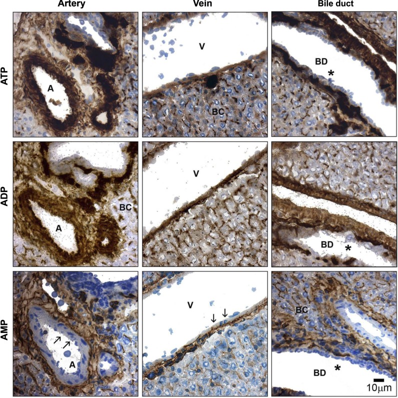
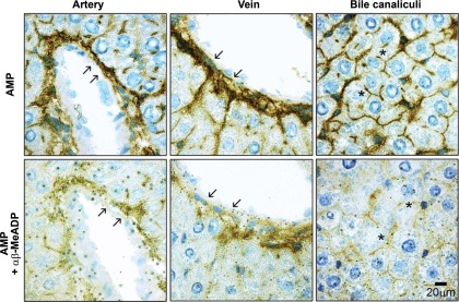
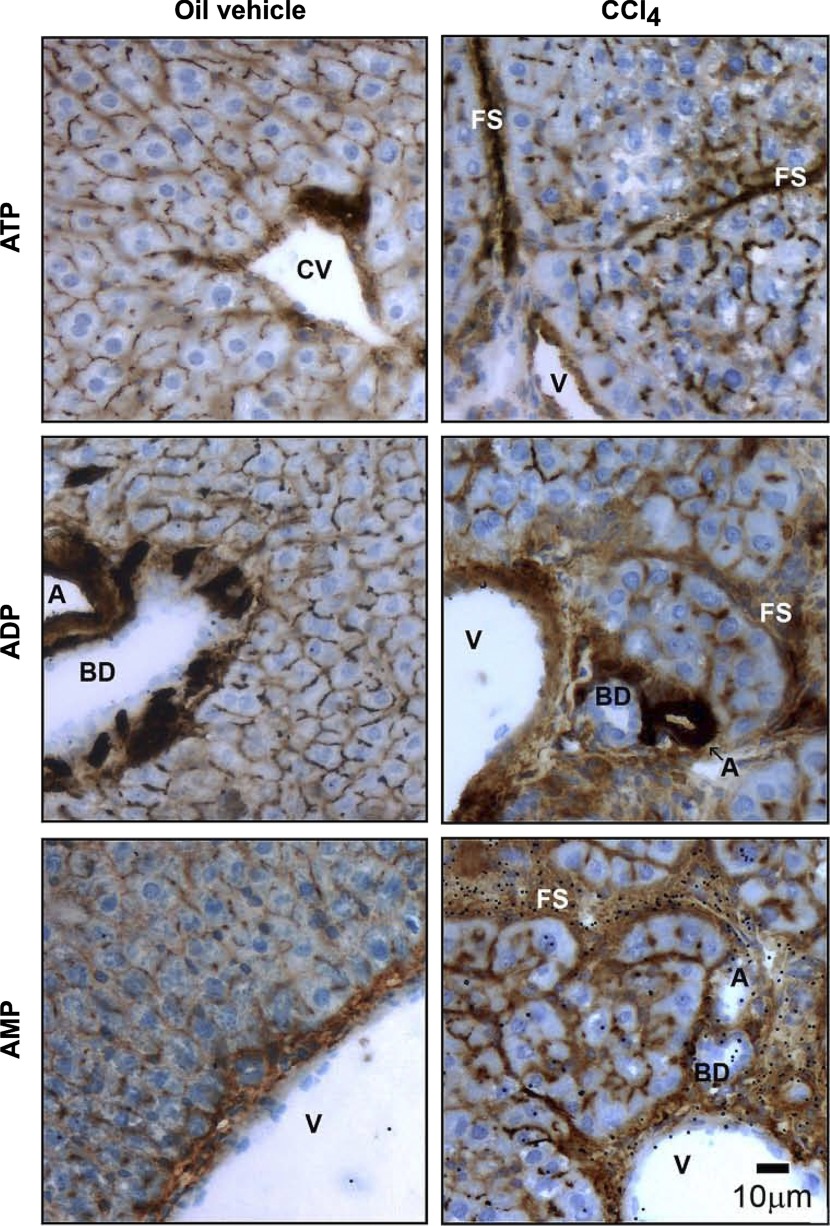
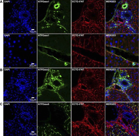
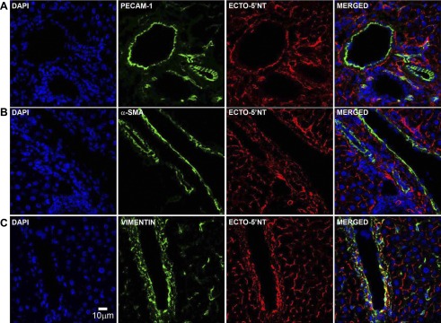
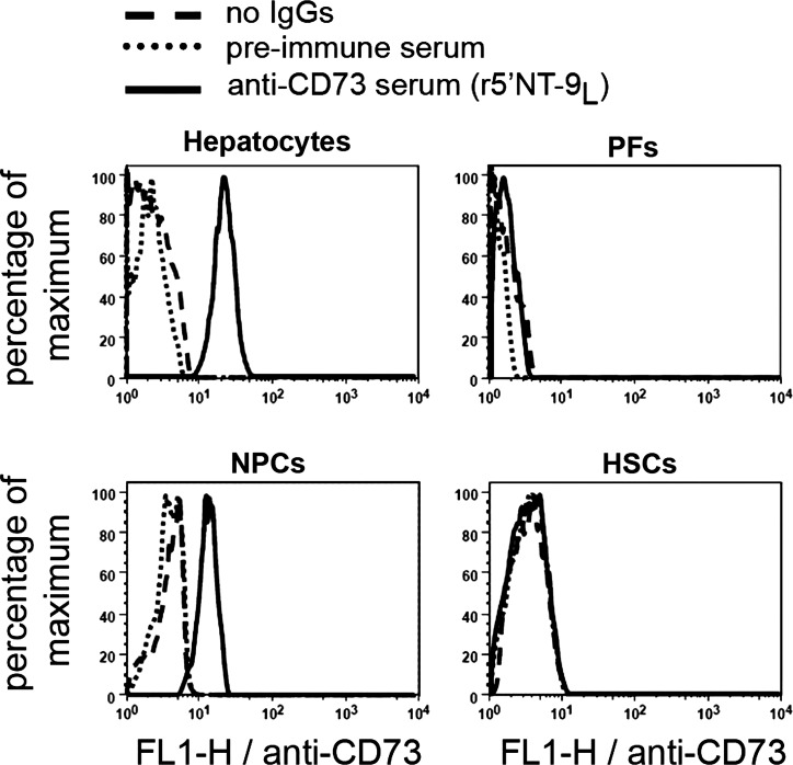
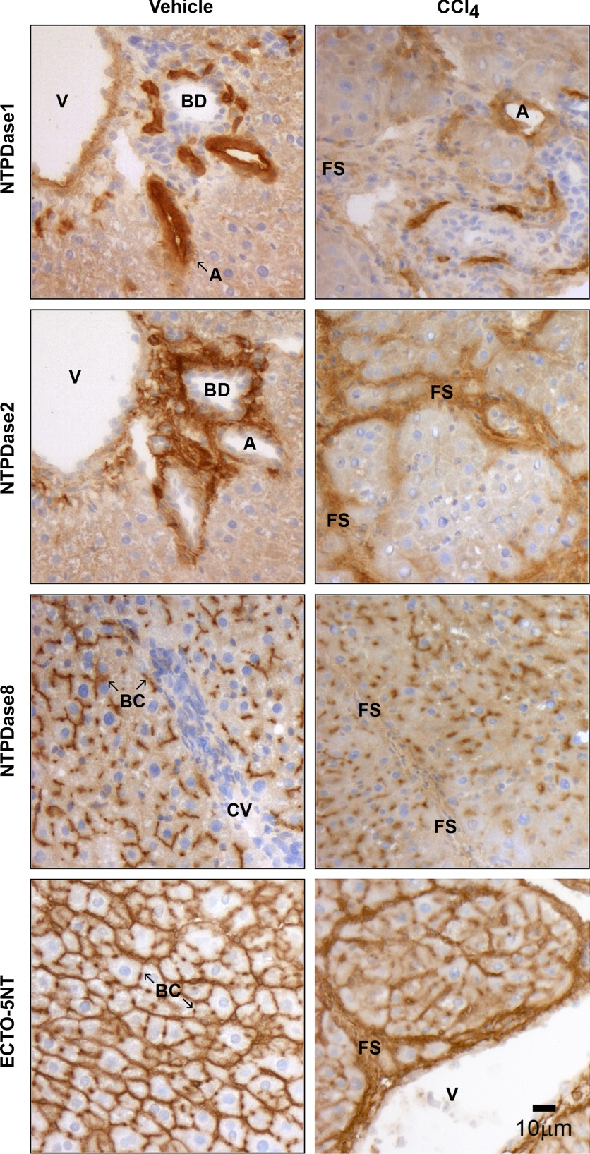
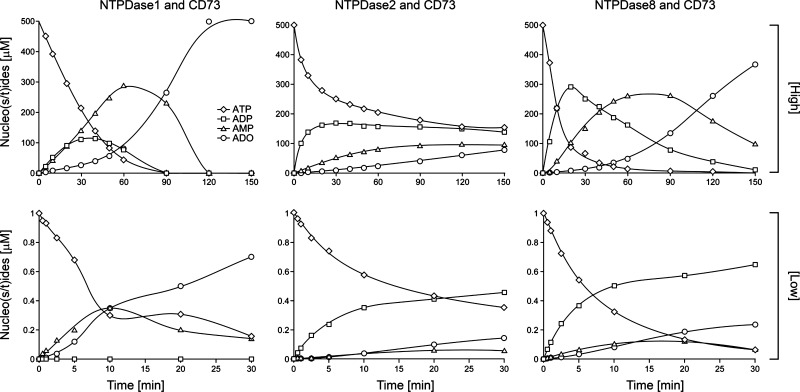
Ectonucleotidases modulate purinergic signaling by hydrolyzing ATP to adenosine. Here we characterized the impact of the cellular distribution of hepatic ectonucleotidases, namely nucleoside triphosphate diphosphohydrolase (NTPDase)1/CD39, NTPDase2/CD39L1, NTPDase8, and ecto-5'-nucleotidase/CD73, and of their specific biochemical properties, on the levels of P1 and P2 receptor agonists, with an emphasis on adenosine-producing CD73. Immunostaining and enzyme histochemistry showed that the distribution of CD73 (protein and AMPase activity) overlaps partially with those of NTPDase1, -2, and -8 (protein levels and ATPase and ADPase activities) in normal rat liver. CD73 is expressed in fibroblastic cells located underneath vascular endothelial cells and smooth muscle cells, which both express NTPDase1, in portal spaces in a distinct fibroblast population next to NTPDase2-positive portal fibroblasts, and in bile canaliculi, together with NTPDase8. In fibrotic rat livers, CD73 protein expression and activity are redistributed but still overlap with the NTPDases mentioned. The ability of the observed combinations of ectonucleotidases to generate adenosine over time was evaluated by reverse-phase HPLC with the recombinant rat enzymes at high "inflammatory" (500 μM) and low "physiological" (1 μM) ATP concentrations. Overall, ATP was rapidly converted to adenosine by the NTPDase1+CD73 combination, but not by the NTPDase2+CD73 combination. In the presence of NTPDase8 and CD73, ATP was sequentially dephosphorylated to the CD73 inhibitor ADP, and then to AMP, thus resulting in a delayed formation of adenosine. In conclusion, the specific cellular cocompartmentalization of CD73 with hepatic NTPDases is not redundant and may lead to the differential activation of P1 and P2 receptors, under normal and fibrotic conditions.


Figures

Fig. 1.
Fig. 1.
Histochemical detection of liver ectonucleotidase activities. Ectonucleotidase activities were located in serial sections of a normal rat liver, using various nucleotides (200 μM) as substrates. All 3 structural components of a classical hepatic triad are depicted, including branches of hepatic artery (A), portal vein (V), and intrahepatic bile duct (BD). ATPase and ADPase catalytic products are mainly detected in all vasculature (arterial > venous), in the connective tissue associated with the portal space, and in the bile canaliculi (BC) (top and middle). The AMP hydrolysis pattern is located in structures similar to those where ATPase and ADPase activities are found, although no signal is observed either in endothelium or in vascular smooth muscle (arrows, bottom). No ectonucleotidase activity is detected in cholangiocytes for any of the substrates tested (asterisks, right). All enzyme histochemical assays were performed in the presence of levamisole (5 mM) to inhibit tissue-nonspecific alkaline phosphatase (TNAP) activity. Scale bar, 10 μm.
Fig. 2.
Fig. 2.
Histochemical detection of liver ecto-5′-nucleotidase activity. Ecto-5′-nucleotidase activity was located in normal rat liver sections with AMP (200 μM) as substrate. Top: prominent signals for hepatic AMPase activity in the connective tissue associated with perivascular areas (arrows), and in bile canaliculi (asterisks). Bottom: ecto-5′-nucleotidase inhibitor α,β-methylene-ADP (αβ-MeADP; 1 mM) reduces AMPase activity in all structures (see arrows and asterisks). All enzyme histochemical assays were performed in the presence of the TNAP inhibitor levamisole (5 mM). Scale bar, 20 μm.
Fig. 3.
Fig. 3.
Histochemical detection of liver ectonucleotidase activities in CCl4-induced hepatic fibrosis model. Ectonucleotidase activities were located in liver sections from vehicle (control) and CCl4-treated rats, using ATP, ADP, or AMP (200 μM) as substrate. All 3 structural components of a classical hepatic triad are depicted: branches of hepatic artery, portal vein, and the intrahepatic bile duct. ATPase, ADPase, and AMPase activities in control animals followed a distribution pattern similar to that described in Fig. 1. In contrast, in fibrotic animals, overlapping signals for ATP, ADP, and AMP hydrolytic products were detected in fibrous bands surrounding hepatic nodules. Moreover, staining for ATPase and ADPase activities was observed in hepatic sinusoids and hepatocyte basolateral membrane, and more prominently, although in a diffuse manner, in bile canaliculi. All enzyme histochemical assays were performed in the presence of the TNAP activity inhibitor levamisole (5 mM). CV, central vein; FS, fibrous septum. Scale bar, 10 μm.
Fig. 4.
Fig. 4.
Specificity of the polyclonal antibodies to rat ecto-5′-nucleotidase. Immunocytochemistry was performed with intact nontransfected (insets) or transfected COS-7 cells with an expression vector encoding rat ecto-5′-nucleotidase. Cells were incubated with either preimmune serum or guinea pig (r5′NT-4c; top) or rabbit (r5′NT-9l; bottom) polyclonal antibody to ecto-5′-nucleotidase. Immunoreactivity is present only in transfected cells incubated with either antibody. Scale bar, 20 μm. Immunoblot analysis of lysates from nontransfected COS-7 cells (ctrl) next to lysates from COS-7 cells transfected with the ecto-5′-nucleotidase expression vector (CD73) shows a single band of ∼66 kDa found exclusively in lysates from transfected cells, again confirming the specificity of both guinea pig r5′NT-4c and rabbit r5′NT-9l sera. A: guinea pig antibody (r5′NT-4c; right) or its preimmune serum (left). B: rabbit antibody (r5′NT-9l; right) or its preimmune serum (left).
Fig. 5.
Fig. 5.
Comparative distribution of liver nucleoside triphosphate diphosphohydrolases (NTPDases) and ecto-5′-nucleotidase (ECTO-5′NT). Double immunofluorescence labeling of NTPDases and ecto-5′-nucleotidase was performed in serial sections of rat liver. A: NTPDase1 staining is associated with the liver vascular network and Kupffer cells. B: NTPDase2 labeling is detected in the connective tissue of portal and septal spaces and perivascular areas. C: NTPDase8 signal is present exclusively in bile canaliculi. AC: ecto-5′-nucleotidase immunostaining is observed in the connective tissue found in portal space or perivascular areas as well as in bile canaliculi. Double staining of NTPDase1 and ecto-5′-nucleotidase reveals that both ectoenzymes are expressed by different but adjacent cell populations in the vasculature, hepatic sinusoids, and hepatic extracellular matrix (A, MERGED). Similarly, NTPDase2 and ecto-5′-nucleotidase proteins are both present at the surface of distinct cell types of hepatic connective tissue (B, MERGED). Both NTPDase8 and ecto-5′-nucleotidase colocalize in bile canaliculi (C, MERGED). Scale bar, 10 μm except for A, bottom, where scale bar is 20 μm. DAPI, 4',6-diamidino-2-phenylindole.
Fig. 6.
Fig. 6.
Comparative distribution of liver ecto-5′-nucleotidase with cell markers. Double immunofluorescence labeling of ecto-5′-nucleotidase and cell markers for vascular endothelial cells [platelet/endothelial cell adhesion molecule-1 (PECAM-1)], myofibroblastic cells (α-smooth muscle actin), and nonmyofibroblastic cells (vimentin) was performed in rat liver sections. Although ecto-5′-nucleotidase immunostaining is clearly distinct from that of PECAM-1 and α-SMA, it partially overlaps with vimentin staining. Scale bar, 10 μm.
Fig. 7.
Fig. 7.
Flow cytometry analysis of ecto-5′-nucleotidase expression in liver primary cell populations. Cell populations were prepared as described in experimental procedures. Ecto-5′-nucleotidase (CD73) was labeled using rabbit polyclonal r5′NT-9l serum. Isolated hepatocytes and nonparenchymal cells (NPCs) are positive for ecto-5′-nucleotidase staining whereas portal fibroblasts (PFs) and hepatic stellate cells (HSCs) are mostly negative.
Fig. 8.
Fig. 8.
Comparative distribution of liver NTPDases and ecto-5′-nucleotidase in CCl4-induced hepatic fibrosis model. Enzyme localization of the ectonucleotidases NTPDase1, -2, -8 and ecto-5′-nucleotidase was determined by immunohistochemistry in liver sections from vehicle (control) and CCl4-treated rats. In control animals, the expression patterns of NTPDase1, -2, -8 and ecto-5′-nucleotidase are similar to those described in Fig. 5. In contrast, in fibrotic animals, NTPDase1 expression is mainly associated with proliferating vascular structures. NTPDase2 expression is almost exclusively detected in perinodular fibrous areas. NTPDase8 protein is only found in bile canaliculi, although with a diffuse distribution. Immunoreactivity to ecto-5′-nucleotidase is localized at the level of both canalicular and basolateral membrane domains in hepatocytes, and prominently in fibrous septa surrounding hepatic fibrotic nodules. Scale bar, 10 μm.
Fig. 9.
Fig. 9.
ATP hydrolysis profile by the different combinations of ecto-5′-nucleotidase with hepatic NTPDases. Hydrolysis products of 500 μM (high) and 1 μM (low) ATP were analyzed over time, as indicated in experimental procedures. At high substrate concentration, ATP is rapidly and completely converted into adenosine in the presence of both NTPDase1 and ecto-5′-nucleotidase (CD73), whereas adenosine is hardly produced by the NTPDase2 + ecto-5′-nucleotidase combination. ATP hydrolysis in the presence of NTPDase8 + ecto-5′-nucleotidase combination generated a transient accumulation of ADP and AMP that resulted in a delayed (compared with the NTPDase1 + ecto-5′-nucleotidase combination) but substantial adenosine production (when compared with the NTPDase2 + ecto-5′-nucleotidase combination). At low substrate level, the incubation of ATP with NTPDase1 + ecto-5′-nucleotidase leads to its rapid dephosphorylation to adenosine without an increase in ADP level. In the presence of either NTPDase2 or NTPDase8 together with ecto-5′-nucleotidase, the accumulation of generated ADP was accompanied by poor AMP production because of limited ADP hydrolysis and thus resulted in a modest adenosine generation by ecto-5′-nucleotidase. ADO, adenosine.

Similar articles

Cited by

References

    1. Airas L, Salmi M, Jalkanen S. Lymphocyte-vascular adhesion protein-2 is a novel 70-kDa molecule involved in lymphocyte adhesion to vascular endothelium. J Immunol 151: 4228–4238, 1993. - PubMed
    1. Bachmann S, Ramasubbu K. Immunohistochemical colocalization of the alpha-subunit of neutrophil NADPH oxidase and ecto-5′-nucleotidase in kidney and liver. Kidney Int 51: 479–482, 1997. - PubMed
    1. Baykov AA, Evtushenko OA, Avaeva SM. A malachite green procedure for orthophosphate determination and its use in alkaline phosphatase-based enzyme immunoassay. Anal Biochem 171: 266–270, 1988. - PubMed
    1. Beldi G, Banz Y, Kroemer A, Sun X, Wu Y, Graubardt N, Rellstab A, Nowak M, Enjyoji K, Li X, Junger WG, Candinas D, Robson SC. Deletion of CD39 on natural killer cells attenuates hepatic ischemia/reperfusion injury in mice. Hepatology 51: 1702–1711, 2010. - PMC - PubMed
    1. Beldi G, Enjyoji K, Wu Y, Miller L, Banz Y, Sun X, Robson SC. The role of purinergic signaling in the liver and in transplantation: effects of extracellular nucleotides on hepatic graft vascular injury, rejection and metabolism. Front Biosci 13: 2588–2603, 2008. - PMC - PubMed

Publication types