. 2011 Dec 15;25(24):2659-73.
doi: 10.1101/gad.174060.111. Epub 2011 Dec 1.

A TRPC5-regulated calcium signaling pathway controls dendrite patterning in the mammalian brain

Affiliations

A TRPC5-regulated calcium signaling pathway controls dendrite patterning in the mammalian brain

Sidharth V Puram et al. Genes Dev. .

Abstract

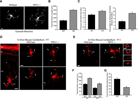
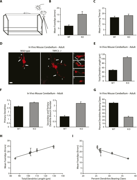
Transient receptor potential (TRP) channels have been implicated as sensors of diverse stimuli in mature neurons. However, developmental roles for TRP channels in the establishment of neuronal connectivity remain largely unexplored. Here, we identify an essential function for TRPC5, a member of the canonical TRP subfamily, in the regulation of dendrite patterning in the mammalian brain. Strikingly, TRPC5 knockout mice harbor long, highly branched granule neuron dendrites with impaired dendritic claw differentiation in the cerebellar cortex. In vivo RNAi analyses suggest that TRPC5 regulates dendrite morphogenesis in the cerebellar cortex in a cell-autonomous manner. Correlating with impaired dendrite patterning in the cerebellar cortex, behavioral analyses reveal that TRPC5 knockout mice have deficits in gait and motor coordination. Finally, we uncover the molecular basis of TRPC5's function in dendrite patterning. We identify the major protein kinase calcium/calmodulin-dependent kinase II β (CaMKIIβ) as a critical effector of TRPC5 function in neurons. Remarkably, TRPC5 forms a complex specifically with CaMKIIβ, but not the closely related kinase CaMKIIα, and thereby induces the CaMKIIβ-dependent phosphorylation of the ubiquitin ligase Cdc20-APC at the centrosome. Accordingly, centrosomal CaMKIIβ signaling mediates the ability of TRPC5 to regulate dendrite morphogenesis in neurons. Our findings define a novel function for TRPC5 that couples calcium signaling to a ubiquitin ligase pathway at the centrosome and thereby orchestrates dendrite patterning and connectivity in the brain.


Figures

Figure 1.
Figure 1.
TRPC5 restricts dendrite growth and elaboration in neurons. (A) Granule neurons transfected with distinct TRPC RNAi plasmids targeting TRPC1, TRPC3, TRPC4, TRPC5, TRPC6, TRPC7, or the control U6 plasmid together with an expression plasmid encoding GFP were subjected to immunocytochemistry 4 d later using the GFP antibody. Representative neurons are shown. In all images of neuronal morphology, arrows and arrowheads indicate dendrites and axons, respectively. TRPC1, TRPC4, and TRPC5 knockdown led to longer, more highly branched dendrites. Bar, 10 μm. (B) Total dendrite length for granule neurons treated as in A was quantified. Total dendrite length was significantly increased in TRPC1, TRPC4, and TRPC5 knockdown neurons compared with control U6-transfected neurons. In contrast, TRPC3, TRPC6, or TRPC7 knockdown had little or no effect on total dendrite length compared with control U6 transfection (ANOVA, P < 0.0001). One-thousand-one-hundred-seventy neurons were measured. Specific values for mean, SEM, and number of cells analyzed for each condition are provided for all experimental results in Supplemental Table 2. A detailed statistics table comparing each shRNA with the others is provided in Supplemental Table 3. The population distribution of total dendrite length for TRPC5 knockdown and control U6-transfected neurons is shown in Supplemental Figure S10. (C) Granule neurons transfected with an expression plasmid encoding TRPC1, TRPC3, TRPC4, TRPC5, TRPC6, TRPC7, or their control vector together with the GFP expression plasmid were analyzed as in A. Expression of TRPC5—but not TRPC1, TRPC3, TRPC4, TRPC6, or TRPC7—substantially reduced dendrite growth and arborization. Bar, 10 μm. (D) Total dendrite length for granule neurons treated as in C was quantified. Total dendrite length was significantly decreased in neurons expressing TRPC5—but not in neurons expressing TRPC1, TRPC3, TRPC4, TRPC6, or TRPC7—compared with control vector-transfected neurons (ANOVA, P < 0.0001). Six-hundred-thirty neurons were measured.
Figure 2.
Figure 2.
TRPC5 is essential for dendrite patterning in the mammalian brain. (A) Granule neurons from TRPC5 wild-type and knockout littermates transfected with GFP were analyzed as in Figure 1A. Representative neurons are shown. Granule neurons from TRPC5 knockout mice had longer, more highly branched dendrites compared with neurons from wild-type littermates. Bar, 10 μm. (B) Granule neurons from TRPC5 wild-type and knockout littermates were treated as in A and subjected to morphometric analysis. Granule neurons from TRPC5 knockout animals had significantly increased total dendrite length compared with neurons from wild-type littermates (t-test, P < 0.0001). One-hundred-sixty-four neurons were measured in six animals (three wild type and three TRPC5 knockout). (C) Granule neurons from TRPC5 wild-type and knockout littermates were treated as in A and subjected to morphometric analysis. Primary dendrite number was modestly increased (t-test, P < 0.005), and secondary and tertiary dendrite branch number was significantly increased in granule neurons from TRPC5 knockout mice compared with wild-type littermates (t-test, P < 0.001). One-hundred-sixty-four neurons were analyzed in six animals (three wild type and three TRPC5 knockout). (D) P7 wild-type and TRPC5 knockout littermate mice were sacrificed, and cerebella were subjected to analysis using a diolistics approach. (Left) Representative wild-type granule neuron, with soma (asterisk) and dendrites (arrows) in the IGL and ascending axon (arrowhead) connecting to the horizontally oriented parallel fibers superficially. Bar, 10 μm. (Right) Representative IGL granule neurons in wild-type and TRPC5 knockout animals are shown. IGL granule neurons in TRPC5 knockout animals had longer, more highly branched dendrites compared with IGL granule neurons in wild-type littermates. Bar, 10 μm. (E) P11 wild-type and TRPC5 knockout littermate mice were sacrificed, and cerebella were analyzed as in D. Representative IGL granule neurons in wild-type and TRPC5 knockout animals are shown. Bar, 10 μm. (Inset) Zoomed view of dendritic tips of individual neurons. Bar, 2.5 μm. Bracket identifies dendritic claws. IGL granule neurons in TRPC5 knockout animals had longer, more highly branched dendrites with fewer dendritic claws compared with IGL granule neurons in wild-type littermates. (F) IGL granule neurons analyzed as in D and E were subjected to morphometric analysis. Total dendrite length was significantly increased in IGL granule neurons in P7 and P11 TRPC5 knockout animals compared with wild-type littermates (ANOVA, P < 0.0001). Three-hundred-seventy-eight neurons were measured in 12 animals (three wild type and three TRPC5 knockout for each age). (G) IGL granule neurons analyzed as in E were subjected to morphometric analysis. The percentage of dendrites bearing claws was significantly reduced in IGL granule neurons in P11 TRPC5 knockout animals compared with wild-type littermates (t-test, P < 0.0001). One-hundred-eighty neurons were analyzed in six animals (three wild type and three TRPC5 knockout).
Figure 3.
Figure 3.
TRPC5 knockout stimulates dendrite growth and arborization in hippocampal neurons. (A) Hippocampal neurons from TRPC5 wild-type and knockout littermates were subjected to immunocytochemistry using the α-tubulin and MAP2 antibody. Representative neurons are shown. TRPC5 knockout neurons had longer, more branched dendrites as compared with neurons from wild-type littermates. Bar, 20 μm. (B) Hippocampal neurons from TRPC5 wild-type and knockout littermates were analyzed as in A and total dendrite length was quantified. Total dendrite length was significantly increased in TRPC5 knockout neurons compared with neurons from wild-type littermates (t-test, P < 0.005). One-hundred neurons were measured in six animals (three wild type and three TRPC5 knockout). (C) Hippocampal neurons from TRPC5 wild-type and knockout littermates were analyzed as in A and primary dendrite number was quantified. Primary dendrite number was not significantly different in TRPC5 knockout neurons compared with neurons from wild-type littermates. One-hundred neurons were analyzed in six animals (three wild type and three TRPC5 knockout). (D) Hippocampal neurons from TRPC5 wild-type and knockout littermates were analyzed as in A and secondary and tertiary dendrite branch number was quantified. Secondary and tertiary dendrite branch number was significantly increased in TRPC5 knockout neurons compared with neurons from wild-type littermates (t-test, P < 0.01). One-hundred neurons were analyzed in six animals (three wild type and three TRPC5 knockout). (E) P7 wild-type and TRPC5 knockout animals were sacrificed and hippocampal sections were analyzed using a diolistics approach as in Figure 2D. Representative CA1 pyramidal neurons in wild-type and TRPC5 knockout animals are shown. CA1 pyramidal neurons in TRPC5 knockout animals had longer, more highly branched dendrites compared with CA1 pyramidal neurons in wild-type littermates. Bar, 25 μm. (F) CA1 pyramidal neurons analyzed as in E were subjected to morphometric analysis. Basolateral dendrite length was modestly increased, and apical and total dendrite length were significantly increased in CA1 pyramidal neurons in TRPC5 knockout animals compared with wild-type littermates (ANOVA, P < 0.0001). Ninety neurons were measured in six animals (three wild type and three TRPC5 knockout). (G) CA1 pyramidal neurons analyzed as in E were subjected to morphometric analysis. Primary dendrite number was modestly increased in CA1 pyramidal neurons in TRPC5 knockout animals compared with wild-type littermates (t-test, P < 0.01). Ninety neurons were measured in six animals (three wild type and three TRPC5 knockout). (H) CA1 pyramidal neurons analyzed as in E were subjected to morphometric analysis. The number of basolateral dendrite branch points was modestly increased, and apical and total dendrite branch points were significantly increased in CA1 pyramidal neurons in TRPC5 knockout animals compared with wild-type littermates (ANOVA, P < 0.0001). Ninety neurons were analyzed in six animals (three wild type and three TRPC5 knockout). (I) P11 wild-type and TRPC5 knockout animals were sacrificed, and hippocampal sections were analyzed using a diolistics approach as in E and subjected to morphometric analysis. Basolateral dendrite length was modestly increased, and apical and total dendrite length were significantly increased in CA1 pyramidal neurons in TRPC5 knockout animals compared with wild-type littermates (ANOVA, P < 0.0001). Ninety neurons were measured in six animals (three wild type and three TRPC5 knockout). (J) CA1 pyramidal neurons analyzed as in I were subjected to morphometric analysis. Primary dendrite number was modestly increased in CA1 pyramidal neurons in TRPC5 knockout animals compared with wild-type littermates (t-test, P < 0.005). Ninety neurons were measured in six animals (three wild type and three TRPC5 knockout). (K) CA1 pyramidal neurons analyzed as in I were subjected to morphometric analysis. The number of basolateral dendrite branch points was modestly increased, and apical and total dendrite branch points were significantly increased in CA1 pyramidal neurons in TRPC5 knockout animals compared with wild-type littermates (ANOVA, P < 0.0001). Ninety neurons were analyzed six animals (three wild type and three TRPC5 knockout).
Figure 4.
Figure 4.
TRPC5 drives dendrite patterning in a cell-autonomous manner in vivo. (A) Lysates of 293T cells transfected with an expression plasmid encoding TRPC5-WT-GFP or TRPC5-RES-GFP together with the TRPC5 RNAi or control U6 plasmid were immunoblotted with the GFP or Actin antibody. The relative density of the TRPC5-GFP band (normalized to Actin) is shown below each lane. (B) Granule neurons transfected with the TRPC5 RNAi or control U6 plasmid together with the expression plasmid encoding TRPC5-WT, TRPC5-RES, or control vector and the GFP expression plasmid were analyzed as in Figure 1A. Expression of TRPC5-RES, but not TRPC5-WT, substantially reduced dendrite growth and arborization compared with control vector in the background of TRPC5 RNAi. Bar, 10 μm. (C) Total dendrite length for granule neurons treated as in B was quantified. Expression of TRPC5-RES, but not TRPC5-WT, significantly reduced total dendrite length compared with control vector in the background of TRPC5 RNAi (ANOVA, P < 0.0001). Three-hundred-sixty neurons were measured. (D) Rat pups electroporated in vivo with a U6-TRPC5i/CMV-GFP RNAi or control U6/CMV-GFP plasmid were sacrificed 5 d after electroporation (P8), and cerebella (Cb) were subjected to immunohistochemistry using the GFP and Calbindin antibody. Representative neurons for each condition are shown. IGL granule neurons in TRPC5 knockdown animals had longer, more highly branched dendrites than IGL granule neurons in control U6 animals. The asterisk indicates process from another neuron. Bar, 10 μm. (E) IGL granule neurons analyzed as in D were subjected to morphometric analysis. Total dendrite length was significantly increased in IGL granule neurons in TRPC5 knockdown animals compared with control U6 animals (ANOVA, P < 0.0001). Two-hundred-forty-six neurons were measured in nine animals (three for each condition). (F) Rat pups electroporated in vivo with the U6-TRPC5i/CMV-GFP RNAi or control U6/CMV-GFP plasmid were sacrificed at P12 and analyzed as in D. Representative IGL granule neurons for each condition are shown. Bar, 10 μm. (Inset) Zoomed view of dendritic tips of individual neurons. Bar, 2.5 μm. Bracket identifies dendritic claws. IGL granule neurons in TRPC5 knockdown animals had longer, more highly branched dendrites with fewer dendritic claws than IGL granule neurons in control U6 animals. (G) IGL granule neurons analyzed as in F were subjected to morphometric analysis. Total dendrite length was significantly increased in IGL granule neurons in TRPC5 knockdown animals compared with control U6 animals (t-test, P < 0.0001). One-hundred-eighty-four neurons were measured in six animals (three control and three knockdown). (H) IGL granule neurons analyzed as in F were subjected to morphometric analysis. The percentage of dendrites bearing claws was significantly decreased in IGL granule neurons in TRPC5 knockdown animals compared with control U6 animals (t-test, P < 0.0001). One-hundred-eighty-four neurons were analyzed in six animals (three control and three knockdown).
Figure 5.
Figure 5.
TRPC5 knockout mice have motor coordination deficits. (A) Schematic of balance beam assay used to assess motor coordination of wild-type and TRPC5 knockout littermates. (B) Number of foot slips (errors) on a narrow (4-mm-wide) balance beam was quantified in wild-type and TRPC5 knockout littermates. Knockout mice had significantly more foot slips than wild-type littermates (t-test, P < 0.01). Sixteen littermates were analyzed (eight wild type and eight TRPC5 knockout). (C) Mean crossing time in wild-type and TRPC5 knockout littermates analyzed as in B was quantified. Wild-type and TRPC5 knockout mice did not have significantly different mean crossing times. Sixteen littermates were analyzed (eight wild type and eight TRPC5 knockout). (D) Adult wild-type and TRPC5 knockout littermate mice were sacrificed, and cerebella were analyzed as in Figure 2D. Representative IGL granule neurons in wild-type and TRPC5 knockout animals are shown. Bar, 10 μm. (Inset) Zoomed view of dendritic tips of individual neurons. Bar, 2.5 μm. The bracket identifies dendritic claws. IGL granule neurons in adult TRPC5 knockout animals had longer, more highly branched dendrites with fewer dendritic claws compared with IGL granule neurons in wild-type littermates. (E) IGL granule neurons analyzed as in D were subjected to morphometric analysis. Total dendrite length was significantly increased in IGL granule neurons in adult TRPC5 knockout animals compared with wild-type littermates (t-test, P < 0.0001). Two-hundred-ninety-one neurons were measured in 10 animals (five wild type and five TRPC5 knockout). (F) IGL granule neurons analyzed as in D were subjected to morphometric analysis. Primary dendrite number was modestly increased (t-test, P < 0.0001), and secondary and tertiary dendrite branch number was significantly increased in adult TRPC5 knockout animals compared with wild-type littermates (t-test, P < 0.0001). Two-hundred-ninety-one neurons were analyzed in 10 animals (five wild type and five TRPC5 knockout). (G) IGL granule neurons analyzed as in D were subjected to morphometric analysis. The percentage of dendrites bearing claws was significantly reduced in IGL granule neurons in adult TRPC5 knockout animals compared with wild-type littermates (t-test, P < 0.0001). Two-hundred-ninety-one neurons were analyzed in 10 animals (five wild type and five TRPC5 knockout). (H) Adult TRPC5 knockout animals tested in behavioral assays were sacrificed, and cerebella were analyzed as in D. Total dendrite length and the number of mean foot slips (errors) were plotted for each individual animal. There was a statistically significant correlation between increased total dendrite length and a greater number of foot slips (errors) (Pearson's correlation coefficient 0.953, P < 0.005). One-hundred-forty-eight neurons were measured in five animals. (I) Adult TRPC5 knockout animals tested in behavioral assays were analyzed as in H. The percentage of dendrites bearing claws and the number of mean foot slips (errors) were plotted for each individual animal. There was a statistically significant correlation between decreased percentage of dendrites bearing claws and a greater number of foot slips (errors) (Pearson's correlation coefficient 0.885, P < 0.01). One-hundred-forty-eight neurons were measured in five animals.
Figure 6.
Figure 6.
TRPC5 promotes centrosomal CaMKIIβ signaling and thereby regulates dendrite morphogenesis. (A) Whole-cerebellar lysates were immunoblotted with the TRPC5, CaMKIIβ, or ERK1/2 antibody. (B) Whole-brain lysates from wild-type and TRPC5 knockout littermates were immunoprecipitated with TRPC5-conjugated agarose beads and immunoblotted with the CaMKIIβ or TRPC5 antibody. Endogenous CaMKIIβ formed a complex with endogenous TRPC5 in wild-type but not TRPC5 knockout neurons. (C) Lysates of cortical neurons were immunoprecipitated with TRPC5- or IgG-conjugated agarose beads and immunoblotted with the CaMKIIβ, CaMKIIα, or TRPC5 antibody. Endogenous CaMKIIβ, but not CaMKIIα, formed a complex with endogenous TRPC5 in neurons. (D) Lysates of 293T cells transfected with the GFP-CaMKIIβ expression plasmid together with an expression plasmid encoding the N terminus (HA-TRPC5-N term), transmembrane domain (HA-TRPC-TM), or C terminus (HA-TRPC5-C term) of TRPC5, or control vector were immunoprecipitated using the HA antibody and immunoblotted with the GFP or HA antibody. (E) Granule neurons transfected with the TRPC5 RNAi or control U6 plasmid together with the expression plasmid encoding TRPC5-RES, TRPC5-RESΔN term, or control vector and the GFP expression plasmid were analyzed as in Figure 1A. TRPC5-RES, but not TRPC5-RESΔN term, significantly reduced granule neuron dendrite length compared with control vector in the background of TRPC5 RNAi (ANOVA, P < 0.0001). Two-hundred-forty neurons were measured. (F) Granule neurons transfected with of one of two different TRPC5 RNAi plasmids (U6-TRPC5i) or the control U6 plasmid together with the GFP expression plasmid were subjected to immunocytochemistry using the GFP or phosphoThr287-CaMKII antibody. Arrows indicate transfected neurons. TRPC5 knockdown substantially reduced the phosphorylation of CaMKIIβ at Thr287 in neurons. Bar, 10 μm. (G) Granule neurons treated as in F were quantified for phosphoThr287-CaMKIIβ signal. The percentage of neurons with phosphoThr287-CaMKII immunoreactivity was significantly reduced in TRPC5 knockdown neurons compared with control U6-transfected neurons (ANOVA, P < 0.005). Two-hundred-seventy-two neurons were analyzed. (H) Granule neurons transfected with the TRPC5 RNAi or control U6 plasmid together with an expression plasmid encoding farnesylated GFP (fGFP) were subjected to immunocytochemistry using the GFP or phosphoSer51-Cdc20 antibody. The percentage of neurons with phosphoSer51-Cdc20 immunoreactivity was significantly reduced in TRPC5 knockdown neurons compared with control U6-transfected neurons (ANOVA, P < 0.005). One-hundred-eighty-four neurons were analyzed. (I) Granule neurons transfected with the expression plasmid encoding TRPC5-WT, TRPC5-ΔN term, or control vector together with the fGFP expression plasmid were analyzed as in H. The percentage of neurons with phosphoSer51-Cdc20 immunoreactivity was significantly increased in TRPC5-WT-expressing neurons, but not TRPC5-ΔN term-expressing neurons, as compared with control vector-transfected neurons (ANOVA, P < 0.0001). Five-hundred-eight-one neurons were analyzed. (J) Granule neurons transfected with the TRPC5 expression plasmid or control vector together with the CaMKIIβ RNAi or control U6 plasmid and the GFP expression plasmid were analyzed as in E. Expression of TRPC5 significantly reduced total dendrite length compared with control. CaMKIIβ RNAi significantly increased total dendrite length in neurons in the presence or absence of TRPC5 expression (ANOVA, P < 0.0001). Three-hundred-sixty neurons were measured. (K) Granule neurons transfected with the Cdc20 RNAi or control U6 plasmid together with the TRPC5-DN expression plasmid or control vector and the GFP expression plasmid were analyzed as in E. Expression of TRPC5-DN significantly increased total dendrite length compared with control. Cdc20 RNAi significantly reduced total dendrite length in neurons in the presence or absence of TRPC5-DN (ANOVA, P < 0.0001). Three-hundred-sixty neurons were measured. (L) Model of TRPC5 regulation of centrosomal CaMKIIβ signaling in the control of dendrite patterning in the mammalian brain.

Similar articles

Cited by

References

    1. Altman J, Bayer S 1997. Development of the cerebellar system: In relation to its evolution, structure, and functions. CRC Press, New York
    1. Baba Y, Hayashi K, Fujii Y, Mizushima A, Watarai H, Wakamori M, Numaga T, Mori Y, Iino M, Hikida M, et al. 2006. Coupling of STIM1 to store-operated Ca2+ entry through its constitutive and inducible movement in the endoplasmic reticulum. Proc Natl Acad Sci 103: 16704–16709 - PMC - PubMed
    1. Bingol B, Schuman EM 2005. Synaptic protein degradation by the ubiquitin proteasome system. Curr Opin Neurobiol 15: 536–541 - PubMed
    1. Bingol B, Schuman EM 2006. Activity-dependent dynamics and sequestration of proteasomes in dendritic spines. Nature 441: 1144–1148 - PubMed
    1. Brewer GJ 1995. Serum-free B27/neurobasal medium supports differentiated growth of neurons from the striatum, substantia nigra, septum, cerebral cortex, cerebellum, and dentate gyrus. J Neurosci Res 42: 674–683 - PubMed

Publication types

Substances