. 2012 Feb 1;185(3):301-10.
doi: 10.1164/rccm.201106-1027OC. Epub 2011 Dec 1.

Cystic fibrosis transmembrane conductance regulator regulates epithelial cell response to Aspergillus and resultant pulmonary inflammation

Affiliations

Cystic fibrosis transmembrane conductance regulator regulates epithelial cell response to Aspergillus and resultant pulmonary inflammation

Neelkamal Chaudhary et al. Am J Respir Crit Care Med. .

Abstract

Rationale: Mutations in the cystic fibrosis transmembrane conductance regulator (CFTR) alter epithelial cell (EC) interactions with multiple microbes, such that dysregulated inflammation and injury occur with airway colonization in people with cystic fibrosis (CF). Aspergillus fumigatus frequently colonizes CF airways, but it has been assumed to be an innocent saprophyte; its potential role as a cause of lung disease is controversial.

Objectives: To study the interactions between Aspergillus and EC, and the role of the fungus in evoking inflammatory responses.

Methods: A. fumigatus expressing green fluorescent protein was developed for in vitro and in vivo models, which used cell lines and mouse tracheal EC.

Measurements and main results: Fungal spores (conidia) are rapidly ingested by ECs derived from bronchial cell lines and murine tracheas, supporting a role for EC in early airway clearance. Bronchial ECs harboring CFTR mutations (ΔF508) or deletion demonstrate impaired uptake and killing of conidia, and ECs with CFTR mutation undergo more conidial-induced apoptosis. Germinated (hyphal) forms of the fungus evoke secretion of inflammatory mediators, with CFTR mutation resulting in increased airway levels of macrophage inflammatory protein 2 and KC, and higher lung monocyte chemotactic protein-1. After A. fumigatus inhalation, CFTR(-/-) mice develop exaggerated lymphocytic inflammation, mucin accumulation, and lung injury.

Conclusions: Data demonstrate a critical role for CFTR in mediating EC responses to A. fumigatus. Results suggest that the fungus elicits aberrant pulmonary inflammation in the setting of CFTR mutation, supporting the potential role of antifungals to halt progressive CF lung disease.


Figures
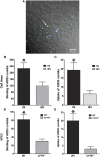
Figure 1.
Figure 1.
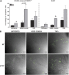
ΔF508 epithelial cell line IB3 and cystic fibrosis transmembrane conductance regulator (CFTR)−/− mouse tracheal epithelial cells (MTEC) demonstrate reduced binding and uptake of green fluorescent protein (GFP)–Af293 conidia. (A) Calcofluor white counterstaining of GFP-expressing conidia distinguishes between internalized conidia (green, arrow) and external conidia (blue). Triplicate wells were prepared for each cell type and six random fields were examined to quantify internalized and external conidia. Average numbers of conidia bound to cell lines (blue) per field are shown in B, and the number of conidia per cell (green) taken up by epithelial cell lines are shown in C. Average numbers of conidia bound and taken up by wild-type (WT) and CFTR−/− MTEC are shown in D and E, respectively. Data represent means ± SEM from three independent experiments. * Statistical significance (two-tailed Student t test; P < 0.05) of conidial uptake by CFTR−/− and ΔF508 IB3 cells.
Figure 2.
Figure 2.
Aspergillus fumigatus (Af293) conidia remain viable inside cystic fibrosis (CF) epithelial cells. Intracellular viability of Af293 conidia inside (A) S9 and (B) IB3 cells was measured by FUN-1 metabolism from green to orange. S9 and IB3 cells grown on 18-mm coverslips were infected with FUN-1–labeled Af293 live conidia (2 × 10⁶) for 7–8 hours at 37°C/5% CO2. Extracellular conidia were counterstained with calcofluor white (blue), and plates were washed and fixed in 1% paraformaldehyde. Conidia inside S9 epithelial cells fluoresced green (A, arrows), whereas germinating conidia that accumulate orange vacuoles were observed more frequently in IB3 cells, indicating fungal metabolic activity (B, arrows), ×63. (C) Green fluorescent protein–Af293 inside mouse tracheal epithelial cells harvested from cystic fibrosis transmembrane conductance regulator deficient mice retained viability after 8 hours, as observed by swelling and germination of intracellular (green) organisms (arrow), ×40.
Figure 3.
Figure 3.
Defective clearance of Aspergillus fumigatus (Af293) conidia by cystic fibrosis transmembrane conductance regulator (CFTR)−/− airways. Representative lung sections with Gomori methenamine silver staining (×100) showing the presence of conidia (arrowheads) in lung parenchyma and in the airways of Af293-challenged (A) wild-type (WT) and (B) CFTR−/− mice at 6 hours. (C) At 24 hours, Af293-challenged WT mice demonstrated no visible conidia or hyphal elements in the airways, whereas hyphal elements were seen (arrowhead) in the large airways of (D) Af293-challenged CFTR−/− mice, ×100.
Figure 4.
Figure 4.
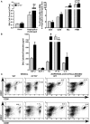
Inflammatory mediators in well-differentiated wild-type (WT) and cystic fibrosis transmembrane conductance regulator (CFTR)−/− mouse tracheal epithelial cells after exposure to Aspergillus fumigatus conidia or germ tubes (GTs). Levels of cytokines and chemokines were measured using Luminex multiplex technology, after exposure to Af293 heat-inactivated conidia/GT (1 × 10⁷; multiplicity of infection of 20:1) or LPS (1 μg/ml) for 24 hours. Only cytokines and chemokines that were differentially measured are shown. Data are representative of two independent experiments. Significance of difference between independent groups of data (mean ± SEM) was analyzed by Student t test (two-tailed); one sample t test was performed using the lower limit of detection for the groups in which the cytokine values were below the assay detection limits (nd, not detected). * P < 0.05 for all comparisons between WT and CFTR−/−. # P < 0.05 for all comparisons of conidia- or GT-treated versus untreated cells in WT and CFTR−/−. IP = IFN-γ– induced protein; MCP = monocyte chemotactic protein; MIP = macrophage inflammatory protein.
Figure 5.
Figure 5.
Epithelial cells harboring ΔF508 mutation or cystic fibrosis transmembrane conductance regulator (CFTR) deletion exhibit increased susceptibility to apoptosis after exposure to Af293 conidia. The percentage of apoptotic S9 (wild-type [WT]) and IB3 (ΔF508) epithelial cell lines treated with Af293 conidia (4 × 10⁶) and staurosporine (STS) (1 μM) (positive control) for 4 hours was measured by flow-cytometry using the (A) fluorescein isothiocyanate anti–poly-ADP ribose polymerase (PARP)-1 antibody and fluorescein isothiocyanate M-30 cytodeath antibody (see Methods). Data (mean ± SEM) represent two independent experiments. *P < 0.05 for comparisons between IB3 and S9 cells, calculated by the Student two-tailed t test. (B) The percentage of apoptotic mouse tracheal epithelial cells (appear green) from WT and CFTR−/− mice was assessed after exposure to Af293 conidia and tumor necrosis factor (TNF)-α (100 ng/ml) (positive control) by fluorescein isothiocyanate M-30 staining (×40).
Figure 6.
Figure 6.
Increased cellular infiltration and elevated levels of inflammatory mediators in bronchoalveolar lavage (BAL) of Af293-challenged cystic fibrosis transmembrane conductance regulator (CFTR)−/− mice. BAL fluid collected after 24 hours from Af293-challenged wild-type (WT) and CFTR−/− mice were evaluated for cellular analysis using flow cytometry (see Methods) and were also analyzed for cytokines and chemokines using the Luminex assay. (A) Higher numbers of macrophages (MΦ) and polymorphonuclear leukocytes (PMN) were observed in BAL of Af293-challenged CFTR−/− mice after 24 hours of conidial exposure than WT mice. (B) Increased levels of macrophage inflammatory protein (MIP)-2 and KC chemokines were also observed in BAL of Af293-challenged CFTR−/−at this time point. Cellular populations were also measured in lung homogenates by flow cytometry at basal levels (untreated) and after 96 hours of conidial exposure in CFTR−/− and WT mice. (C) Higher percentage of T cells (CD3+/CD4+, CD3+/CD8+) were present in the lungs of CFTR−/− mice recovered 96 hours after exposure to Af293 conidia than WT mice. The data are representative of two independent experiments, with BAL and lung cells (pooled) from three mice per condition. Total of 10,000 events were analyzed for flow cytometry data. Significance between independent groups of data was analyzed by Student t test (two-tailed). * P < 0.05, comparison of Af293-challenged WT and CFTR−/−. # P < 0.05, comparison of Af293-challenged WT and CFTR−/− versus basal levels. IP = IFN-γ–induced protein; MCP = monocyte chemotactic protein.
Figure 7.
Figure 7.
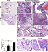
Lung sections stained with hematoxylin and eosin (A–H) and alcian blue (J and K) from wild-type (WT) and cystic fibrosis transmembrane conductance regulator (CFTR)−/− mice challenged with Af293 conidia, observed after 72 hours at indicated magnification. (A) Lung sections from unchallenged WT and (D) CFTR−/− mice showed no basal differences in cellular infiltration and inflammation. (B) Lung sections (×20 magnification) from WT mice challenged with Af293 demonstrated acute inflammation in small airways at adjacent alveoli, bronchioles, and alveolar spaces (AS). (C) Enhanced (×100) magnification of boxed area in B showed intraluminal acute inflammation in small airways without epithelial necrosis or fibrin deposition. (E) Lung sections (×20 magnification) from Af293-infected CFTR−/− mice showed prominent fibrin (F) deposition in the areas of acute inflammation. (F) Enhanced (×100) magnification of boxed region in (E) showed fibrin (F) deposition in the bronchioles with loss of respiratory epithelium (LRE). (G) Prominent intraluminal mucin (M) was present in the bronchiole shown (×20) with (H) magnified view at (×100) of boxed region showing acute inflammation (AI), with accumulated mucin, without epithelial necrosis. (I) Lung inflammatory score was higher in Af293-challenged CFTR−/−, performed using blinded scoring in lung sections (n = 2–3) at 24 and 72 hours. The scoring for the lung inflammatory changes was ranked from 0–4. This scoring parameters included the following: 0, no inflammation around the bronchioles and in alveolar spaces; 1, chronic inflammation around bronchioles; 2, acute and chronic inflammation around bronchioles, and acute inflammation in bronchiolar lumens; 3, acute and chronic inflammation around bronchioles, and acute inflammation in bronchiolar lumens and scattered airspaces; and 4, acute and chronic inflammation around bronchioles, acute inflammation in bronchiolar lumens, epithelial necrosis, and extensive fibrin deposition. (J and K) Representative lung sections (×40) stained with alcian blue demonstrating increased abundance of mucin in airways of (K) Af293-challenged CFTR−/− mice than WT (J).
Figure 8.
Figure 8.
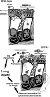
Schematic representation of early events occurring after exposure of wild-type and cystic fibrosis transmembrane conductance regulator (CFTR)−/− epithelial cells (EC) to conidia. Conidia are normally internalized and inactivated by ECs, without evoking secretion of inflammatory mediators (upper panel). In the presence of CFTR mutations, conidia induce increased EC apoptosis. Inefficient conidial uptake and killing results in more luminal conidial germination to hyphal cells, which subsequently trigger increased production of specific inflammatory mediators (e.g., monocyte chemotactic protein-1), resultant dysregulated cellular inflammation.

Similar articles

Cited by

References

    1. Lyczak JB, Cannon CL, Pier GB. Lung infections associated with cystic fibrosis. Clin Microbiol Rev 2002;15:194–222. - PMC - PubMed
    1. Pier GB, Grout M, Zaidi TS. Cystic fibrosis transmembrane conductance regulator is an epithelial cell receptor for clearance of Pseudomonas aeruginosa from the lung. Proc Natl Acad Sci USA 1997;94:12088–12093. - PMC - PubMed
    1. Gadjeva M, Paradis-Bleau C, Priebe GP, Fichorova R, Pier GB. Caveolin-1 modifies the immunity to Pseudomonas aeruginosa. J Immunol 2010;184:296–302. - PMC - PubMed
    1. Teichgraber V, Ulrich M, Endlich N, Riethmuller J, Wilker B, De Oliveira-Munding CC, van Heeckeren AM, Barr ML, von Kurthy G, Schmid KW, et al. . Ceramide accumulation mediates inflammation, cell death and infection susceptibility in cystic fibrosis. Nat Med 2008;14:382–391. - PubMed
    1. Zhang Y, Li X, Grassme H, Doring G, Gulbins E. Alterations in ceramide concentration and pH determine the release of reactive oxygen species by CFTR-deficient macrophages on infection. J Immunol 2010;184:5104–5111. - PubMed

Publication types

MeSH terms

Substances