. 2010 Apr;17(2):29-39.

Computed Tomography (CT) Imaging of Injuries from Blunt Abdominal Trauma: A Pictorial Essay

Affiliations

Computed Tomography (CT) Imaging of Injuries from Blunt Abdominal Trauma: A Pictorial Essay

Radhiana Hassan et al. Malays J Med Sci. 2010 Apr.

Abstract

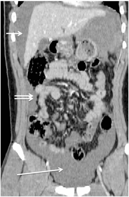
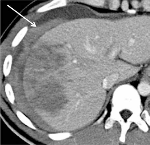
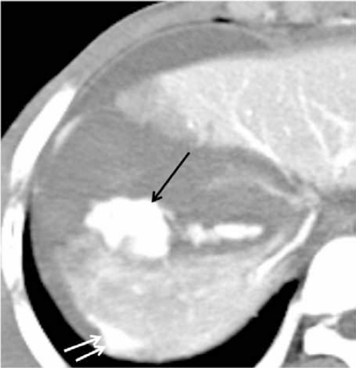
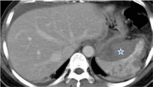
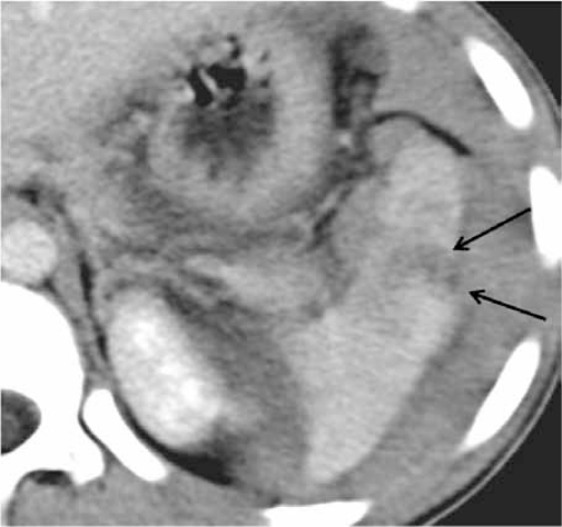
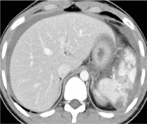
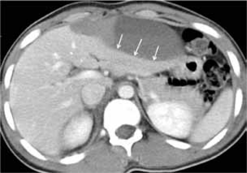
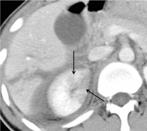
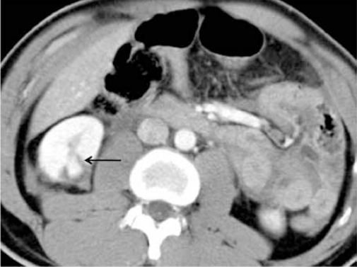
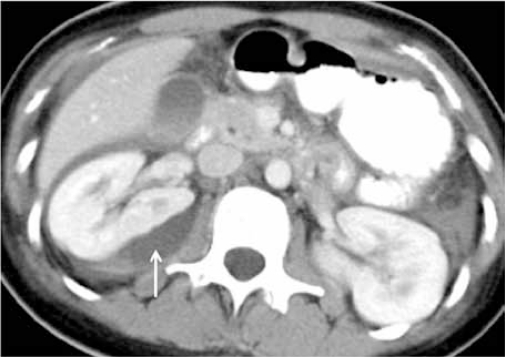
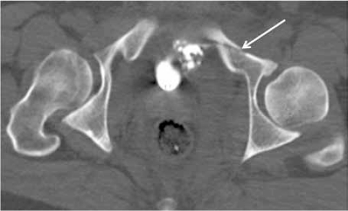
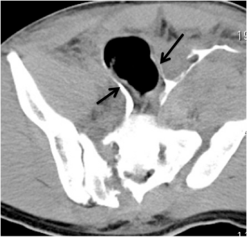
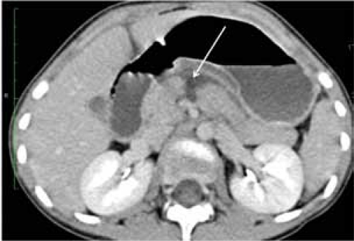
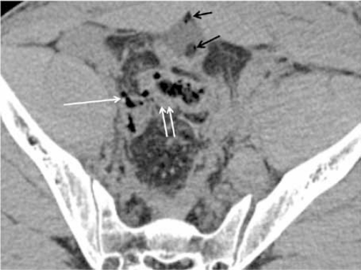
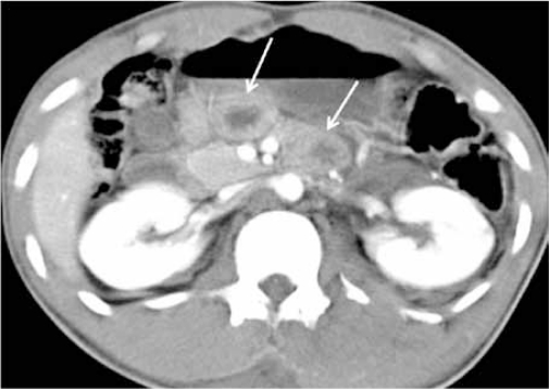
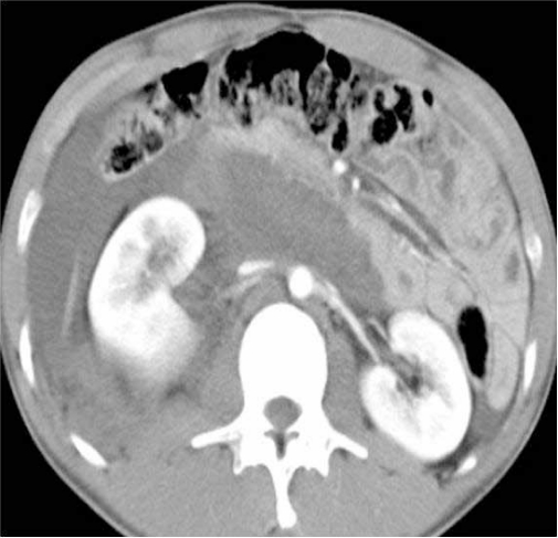
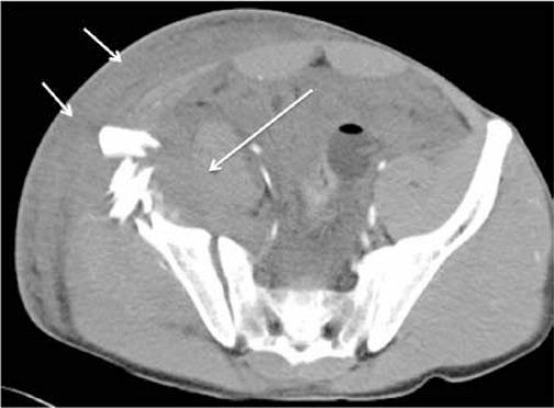
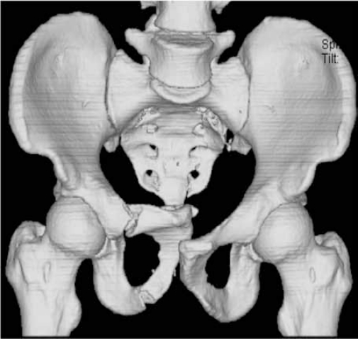
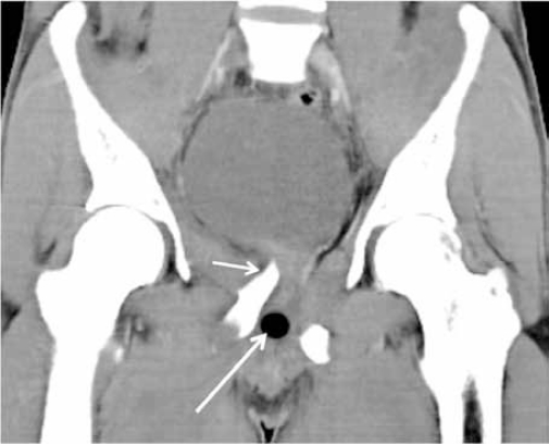
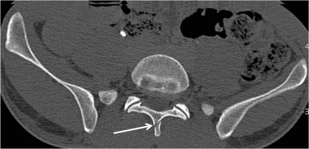
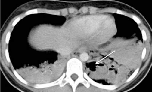
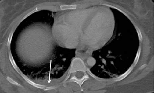
Blunt abdominal trauma can cause multiple internal injuries. However, these injuries are often difficult to accurately evaluate, particularly in the presence of more obvious external injuries. Computed tomography (CT) imaging is currently used to assess clinically stable patients with blunt abdominal trauma. CT can provide a rapid and accurate appraisal of the abdominal viscera, retroperitoneum and abdominal wall, as well as a limited assessment of the lower thoracic region and bony pelvis. This paper presents examples of various injuries in trauma patients depicted in abdominal CT images. We hope these images provide a resource for radiologists, surgeons and medical officers, as well as a learning tool for medical students.

Keywords: blunt abdominal trauma; computed tomography; injuries; medical sciences.


Figures

Figure 1a:
Figure 1a:
CT coronal MPR in 18-year-old boy whose motorbike skidded. He had a Grade V splenic injury (images not shown). Splenectomy was performed and about 2 litres of haemoperitoneum was noted intraoperatively. This image demonstrate the possible pathway of blood flow, from the splenic injury to perihepatic (single arrow) regions and passes down the right paracolic gutter (double arrows) to the pelvic cavity (long arrow).
Figure 1b:
Figure 1b:
CT scan showing haemoperitoneum from liver injury in a 23-year-old man who was involved in a motor vehicle accident (MVA). The ‘sentinel clot’ sign is seen as a high-attenuation collection adjacent to the liver surface (arrow). Liver injury was confirmed surgically with estimated blood loss of 3 litres.
Figure 1c:
Figure 1c:
CT scan demonstrating active haemorrhage in a 20-year-old man following MVA. CT shows contrast extravasation (long arrows) and pooling of the extravasated contrast in the dependant area (short arrows). This patient died 2 days after surgery from excessive blood loss.
Figure 2a:
Figure 2a:
CT scan of a 32-year-old man following MVA showing splenic injury. Subcapsular haematoma (star) appears as a region of low attenuation that compresses the normal splenic parenchyma. Note also multiple lacerations of the spleen. Splenectomy was performed in this patient.
Figure 2b:
Figure 2b:
CT scan showing splenic laceration in a 13-year-old boy, a pillion rider of a skidded motorbike. Splenic laceration is seen as irregular, linear region of low attenuation (arrows). A 4-cm laceration was identified at the tip of the spleen during surgery and splenectomy was performed.
Figure 2c:
Figure 2c:
CT scan demonstrating a shattered spleen in a 21-year-old male motorcyclist following MVA. Multiple hypodense areas that connect to the visceral surfaces are shown. This patient had failed conservative treatment and splenectomy was performed 2 days following the trauma which confirmed the CT findings of shattered spleen.
Figure 3a:
Figure 3a:
CT scan of liver injury in a 48-year-old man with MVA. CT demonstrates a subcapsular haematoma that appears as a hypodense collection, compressing on the underlying liver parenchyma (arrows).
Figure 3b:
Figure 3b:
CT scan of liver injury in a 23-year-old man with MVA. Liver laceration is shown on CT as a non-enhancing irregular, linear low attenuation area (arrow) with associated intraparenchymal haematoma (star), which appears as a region of decrease attenuation compared to the rest of the enhanced liver parenchyma. He was managed surgically.
Figure 4a:
Figure 4a:
CT scan of renal injury in a 20-year-old man with MVA. A right contusion that appears as a focal patchy area of decreased enhancement (arrows) was observed on CT. A liver laceration is present adjacent to the kidney. He was managed conservatively with an uneventful recovery.
Figure 4b:
Figure 4b:
CT of renal laceration in a 32-year-old man with MVA. The right renal lacerations are shown as iregular, linear low attenuation areas within the parenchyma (arrow), which does not involve the collecting systems. He was managed conservatively with an uneventful recovery.
Figure 4c:
Figure 4c:
CT scan of renal injury in a 17-year-old girl with MVA. Subcapsular haematoma of the right kidney (arrows) appears on CT as a superficial, crescentic, low attnuation area that compresses the adjacent renal parenchyma. She was managed conservatively with uneventful recovery.
Figure 5a:
Figure 5a:
CT cystogram of urinary bladder trauma in a 28-year-old man following MVA. There is extravasation of contrast (arrow) in the perivesical fat indicating an extraperitoneal bladder rupture.
Figure 5b:
Figure 5b:
CT scan showing fracture of the pelvic bone. A lower scan of the same patient in Figure 5a shows the pelvic fracture (arrow). He was managed conservatively with uneventful recovery.
Figure 5c:
Figure 5c:
CT scan of urinary bladder injury in a 26-year-old man with industrial accident. He fell and was run over by a tractor. This image shows extravasation of contrast from a urinary bladder injury, which outline the bowel loops (arrows). This indicates an intraperitoneal bladder rupture. Note fracture on the right side of the sacrum and diasthesis of left sacroilliac joint. Urinary bladder perforation at 2 sites with about 1 litre of haemoperitoneum was confirmed at surgery.
Figure 6:
Figure 6:
CT scan of pancreatic transection in a 9-year-old girl with ‘bicycle-handle’ injury. Diagnosis was delayed and CT scan performed 2 days after the incident showed a total transection of the body of pancreas (arrow). This was later complicated by a pseudocyst formation that required a percutaneous drainage.
Figure 7a:
Figure 7a:
CT scan of perforated bowel in a 26-year-old man with MVA. Note subtle extraluminal air (single white arrows) with focal bowel wall thickening (double white arrows) at the rectosigmoid region that was missed on initial review of the CT images. Also note air pockets in the urinary bladder (black arrows). Urinary bladder perforation and transection at the rectosigmoid junction were detected intraoperatively.
Figure 7b:
Figure 7b:
CT scan of bowel injury in a 23-year-old lorry driver with MVA. CT scan showed focal small bowel thickening (arrows) but no free air was identified. Small bowel perforation was found intraoperatively.
Figure 8a:
Figure 8a:
CT scan of retroperitoneal injury in in a 23-year-old man with MVA. CT shows an anterior displacement of the right kidney by a retroperitoneal haemorrhage. Both kidneys are otherwise intact. He was managed conservatively with uneventful recovery.
Figure 8b:
Figure 8b:
CT scan of the same patient in Figure 8a showing the soft tissue injury. There is haematoma and thickening of the abdominal wall (short arrows). The soft tissue injury is extensive involving the right iliopsoas muscle (long arrows) and extends inferiorly to the high region, which compresses the right femoral artery and vein (images are not shown). Note also comminuted fractures of the right iliac bone.
Figure 9a:
Figure 9a:
CT scan showing fracture of the pelvic bone in a 29-year-old lorry driver following MVA. This volume-rendered CT coronal MPR image clearly depicts fracture of the right public rami with displacement of the fractured fragments.
Figure 9b:
Figure 9b:
CT demonstrating soft tissue injury associated with pelvic fracture. A coronal MPR CT image in soft tissue window of the same patient in Figure 9a showed the fractured fragment (short arrow) compressing at the base of the urinary bladder. Note the mal-positioned Foley’s catheter balloon within the urethra (long arrow). Urethrogram demonstrated a membranous urethral injury.
Figure 10:
Figure 10:
CT scan of spine fracture in a 29-year-old man who fell from height at the workplace. He complained of pain at the lumbar region. CT scan shows no-intra abdominal injury but demonstrated a subtle of L5 spinous process (arrow), which was missed on the plain radiograph. He was managed conservatively with uneventful recovery.
Figure 11a:
Figure 11a:
CT of a 15-year-old boy with MVA. The limited evaluation of the lung bases reveals bilateral lung contusions with a left lung laceration (arrow). He sustained a Grade 1 liver injury (not shown) and was managed conservatively.
Figure 11b:
Figure 11b:
CT evaluation of the lower thoracic region in a 25-year-old man with MVA revealed fracture of right posterior rib (arrow) with associated pleural effusion, possibly a haemothorax.

References

    1. Federle MP, Goldberg HI, Kaiser JA, Moss AA, Jeffrey RB, Mail JC. Evaluation of abdominal trauma by computed tomography. Radiology. 1981;138:637–644. - PubMed
    1. Shuman WP. CT of blunt abdominal trauma. Radiology. 1997;205:297–306. - PubMed
    1. Rhea JT. The frequency and significance of thoracic injuries detected on abdominal CT scans of multiple trauma patients. J Trauma. 1989;29(4):502–505. - PubMed
    1. Hoff WS, Holevar M, Nagy KK, Patterson L, Young JS, Arrillaga A, et al. Practice management guidelines for the evaluation of blunt abdominal trauma: the EAST practice management guidelines work group. J Trauma. 2002;53:602–615. - PubMed
    1. Ferrada R, Rivera D, Ferrada P. In: Blunt Abdominal Trauma. General Surgery. 2nd ed. Bland KI, Sarr MG, Buchler MW, Csendes A, et al., editors. London: Springer; 2009. pp. 87–96.

LinkOut - more resources