

Computed tomography of blunt spleen injury: a pictorial review
- PMID: 22135575
- PMCID: PMC3216201
Computed tomography of blunt spleen injury: a pictorial review
Abstract
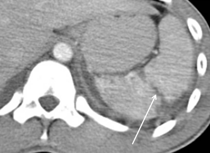
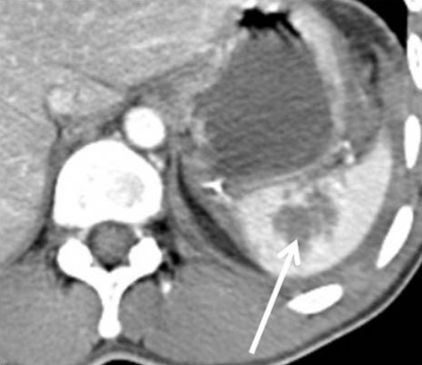
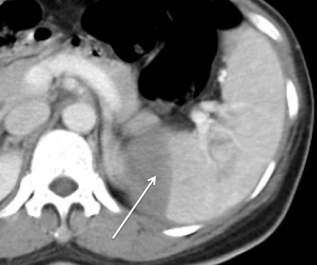
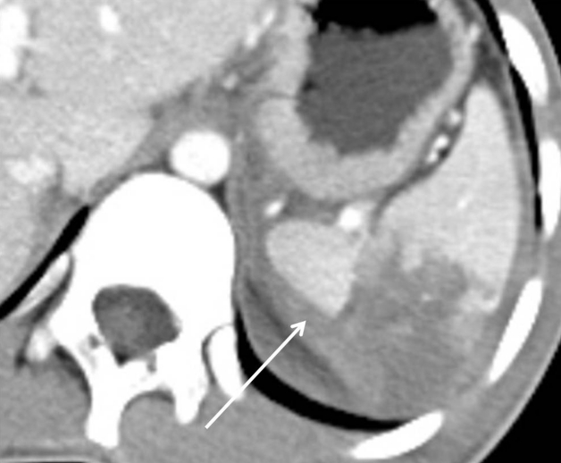
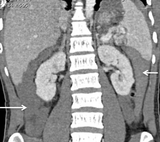
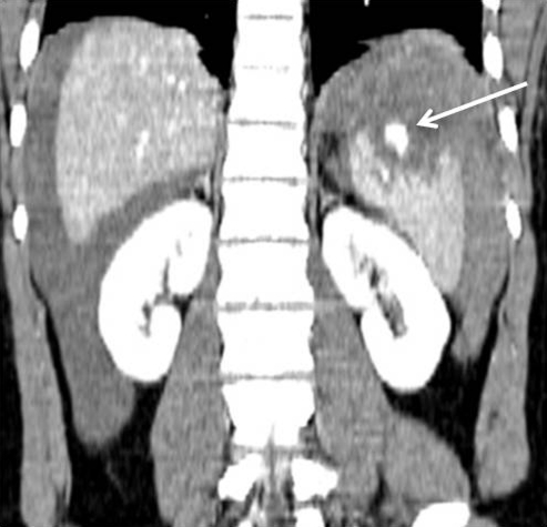
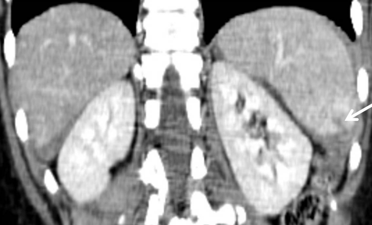
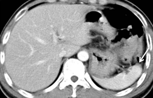
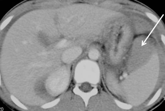
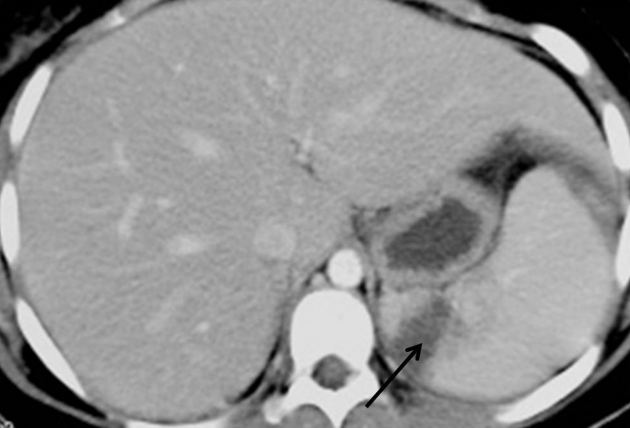
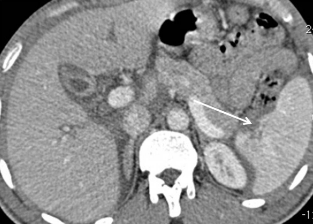
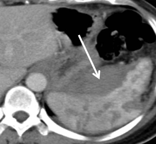
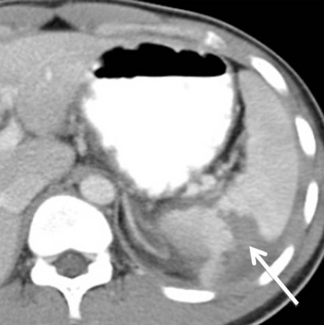
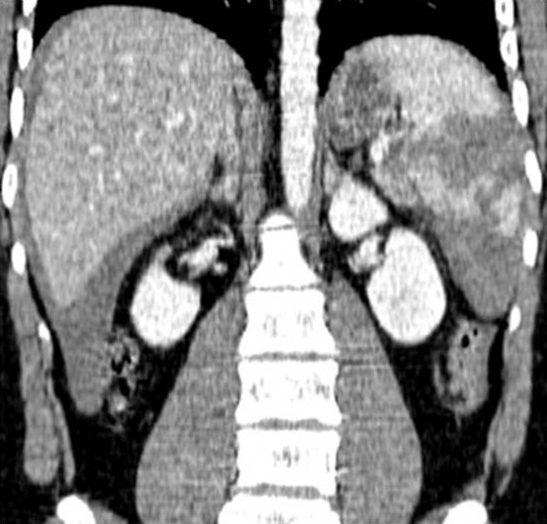
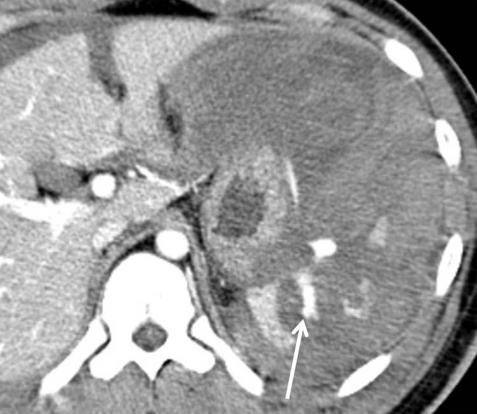
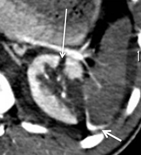
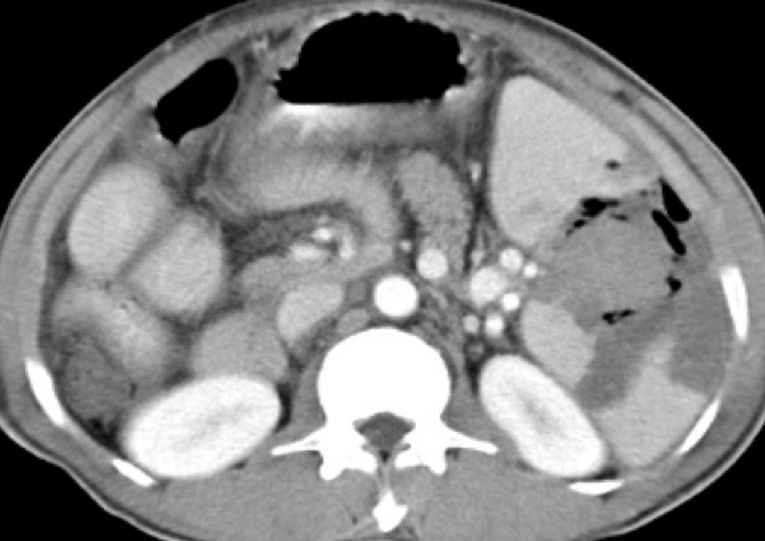
The spleen is one of the organs most frequently injured in blunt abdominal trauma. Computed tomography (CT) scanning can accurately detect splenic injury and is currently the imaging modality of choice in assessing clinically stable patients with blunt abdominal trauma. The CT features of spleen injury include lacerations, subcapsular or parenchymal haematomas, active haemorrhage, and vascular injuries. We present a pictorial review of the spectrum of CT findings for blunt splenic injuries. This article will be a useful reference for radiologists and surgeons as CT scan is widely used for the assessment of splenic injuries and contributes to the current trend towards nonsurgical management of this injury.
Keywords: abdomen; blunt injuries; computed tomography; medical imaging; spleen; trauma.
Figures

Similar articles
-
Computed tomography (CT) in blunt liver injury: a pictorial essay.Med J Malaysia. 2010 Dec;65(4):319-25. Med J Malaysia. 2010. PMID: 21901958
-
CT in blunt liver trauma.Radiographics. 2005 Jan-Feb;25(1):87-104. doi: 10.1148/rg.251045079. Radiographics. 2005. PMID: 15653589 Review.
-
Educational Review of Predictive Value and Findings of Computed Tomography Scan in Diagnosing Bowel and Mesenteric Injuries After Blunt Trauma: Correlation With Trauma Surgery Findings in 163 Patients.Can Assoc Radiol J. 2017 Aug;68(3):276-285. doi: 10.1016/j.carj.2016.07.003. Epub 2017 Jan 23. Can Assoc Radiol J. 2017. PMID: 28126266
-
Blunt splenic trauma: delayed-phase CT for differentiation of active hemorrhage from contained vascular injury in patients.Radiology. 2007 Apr;243(1):88-95. doi: 10.1148/radiol.2431060376. Epub 2007 Feb 9. Radiology. 2007. PMID: 17293574
-
CT of blunt splenic injuries: what the trauma team wants to know from the radiologist.Clin Radiol. 2019 Dec;74(12):903-911. doi: 10.1016/j.crad.2019.07.017. Epub 2019 Aug 28. Clin Radiol. 2019. PMID: 31471062 Review.
Cited by
-
Detection of post-traumatic abdominal pseudoaneurysms by CEUS and CT: A prospective comparative global study (the PseAn study)-study protocol.Front Surg. 2023 Feb 20;10:1124087. doi: 10.3389/fsurg.2023.1124087. eCollection 2023. Front Surg. 2023. PMID: 36891548 Free PMC article.
-
Assessment of blunt splenic trauma: Which imaging scoring system is superior?J Res Med Sci. 2018 Mar 27;23:29. doi: 10.4103/jrms.JRMS_875_17. eCollection 2018. J Res Med Sci. 2018. PMID: 29692826 Free PMC article.
-
Spontaneous splenic rupture in a neonate: a case report and literature review.Emerg Radiol. 2024 Feb;31(1):117-122. doi: 10.1007/s10140-024-02199-0. Epub 2024 Jan 17. Emerg Radiol. 2024. PMID: 38231379 Review.
-
Automated Spleen Injury Detection Using 3D Active Contours and Machine Learning.Entropy (Basel). 2021 Mar 24;23(4):382. doi: 10.3390/e23040382. Entropy (Basel). 2021. PMID: 33804831 Free PMC article.
-
Ultrasonography of the healing process during a 3-month follow-up after a splenic injury.Ultrasonography. 2015 Jul;34(3):226-30. doi: 10.14366/usg.14057. Epub 2014 Dec 30. Ultrasonography. 2015. PMID: 25623053 Free PMC article.
References
-
- Kailidou E, Pikoulis E, Katsiva V, Karavokyros IG, Athanassopoulou A, Papakostantinou I, et al. Contrast-enhanced spiral CT evaluation of blunt abdominal trauma. JBR-BTR. 2005;88(2):61–65. - PubMed
-
- Yao DC, Jeffrey RB, Jr, Mirvis SE, Weekes A, Federle MP, Kim C, et al. Using contrast-enhanced helical CT to visualize arterial extravasation after blunt abdominal trauma: Incidence and organ distribution. AJR Am J Roentgenol. 2002;178(1):17–20. - PubMed
-
- Hoff WS, Holevar M, Nagy KK, Patterson L, Young JS, Arrillaga A, et al. Practice management guidelines for the evaluation of blunt abdominal trauma: The East practice management guidelines work group. J Trauma. 2002;53(3):602–615. - PubMed
-
- Becker CD, Mentha G, Terrier F. Blunt abdominal trauma in adults: Tole of CT in the diagnosis and management of visceral injuries. Part 1: Liver and spleen. Eur Radiol. 1998;8(4):553–562. - PubMed
-
- Fang JF, Wong YC, Lin BC, Hsu YP, Chen MF. Usefulness of multidetector computed tomography for the initial assessment of blunt abdominal trauma patients. World J Surg. 2006;30(2):176–182. - PubMed
LinkOut - more resources
Full Text Sources